

XMRV accelerates cellular proliferation, transformational activity, and invasiveness of prostate cancer cells by downregulating p27(Kip1)
- PMID: 21932423
- PMCID: PMC3275676
- DOI: 10.1002/pros.21491
XMRV accelerates cellular proliferation, transformational activity, and invasiveness of prostate cancer cells by downregulating p27(Kip1)
Abstract
Background: Xenotropic murine leukemia virus-related retrovirus (XMRV) is a recently discovered gammaretrovirus that was originally detected in prostate tumors. However, a causal relationship between XMRV and prostate cancer remains controversial due to conflicting reports on its etiologic occurrence. Even though gammaretroviruses are known to induce cancer in animals, a mechanism for XMRV-induced carcinogenesis remains unknown. Several mechanisms including insertional mutagenesis, proinflammatory effects, oncogenic viral proteins, immune suppression, and altered epithelial/stromal interactions have been proposed for a role of XMRV in prostate cancer. However, biochemical data supporting any of these mechanisms are lacking. Therefore, our aim was to evaluate a potential role of XMRV in prostate carcinogenesis.
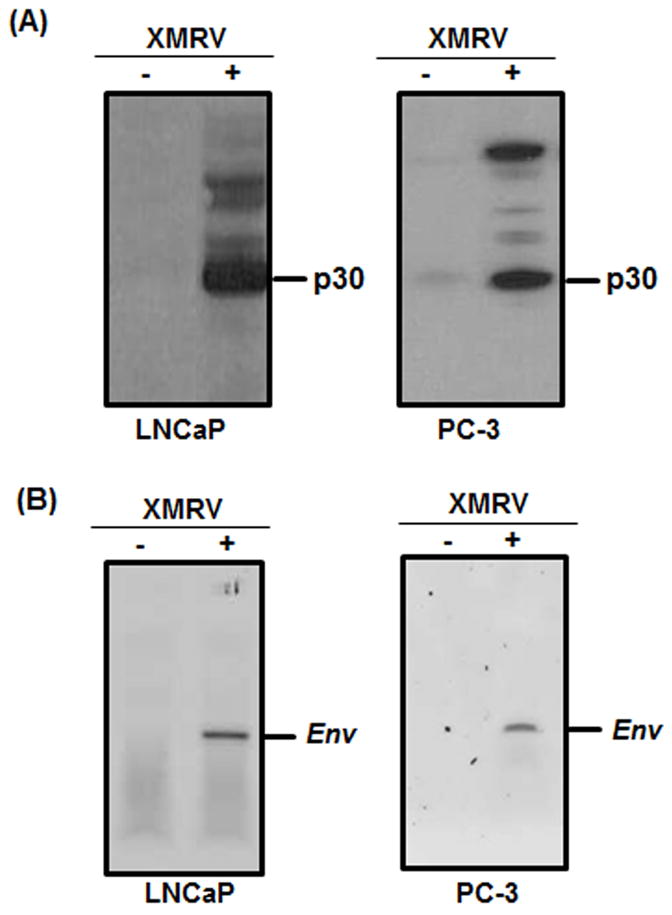
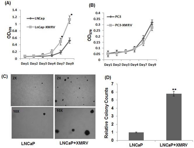
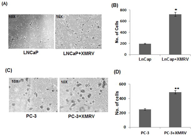
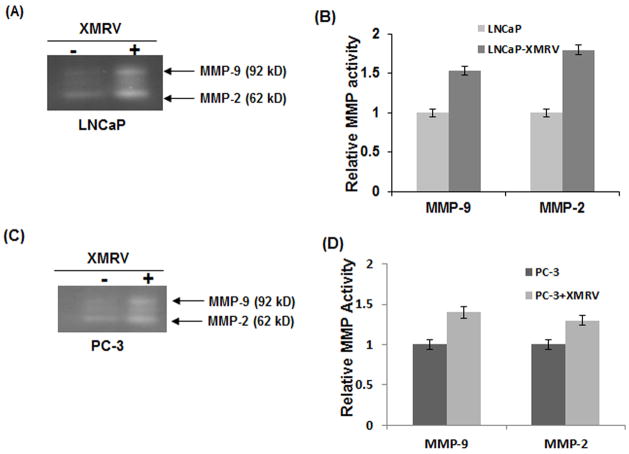
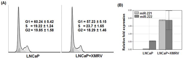
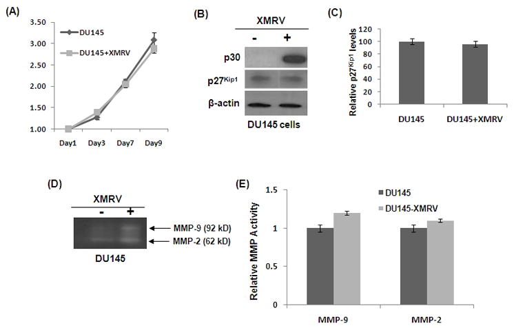
Methods: Growth kinetics of prostate cancer cells are conducted by MTT assay. In vitro transformation and invasion was carried out by soft agar colony formation, and Matrigel cell invasion assay, respectively. p27(Kip1) expression was determined by Western blot and MMP activation was evaluated by gelatin-zymography. Up-regulation of miR221 and miR222 expression was examined by real-time PCR.
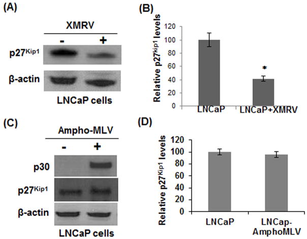
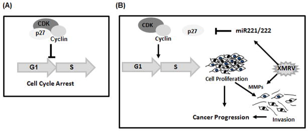
Results: We demonstrate that XMRV infection can accelerate cellular proliferation, enhance transformation, and increase invasiveness of slow growing prostate cancer cells. The molecular basis of these viral induced activities is mediated by the downregulation of cyclin/cyclin dependent kinase inhibitor p27(Kip1) . Downstream analyses illustrated that XMRV infection upregulates miR221 and miR222 expression that target p27(Kip1) mRNA.
Conclusions: We propose that downregulation of p27(Kip1) by XMRV infection facilitates transition of G1 to S, thereby accelerates growth of prostate cancer cells. Our findings implicate that if XMRV is present in humans, then under appropriate cellular microenvironment it may serve as a cofactor to promote cancer progression in the prostate.
Copyright © 2011 Wiley Periodicals, Inc.
Figures
Similar articles
-
miR-196a-mediated downregulation of p27kip1 protein promotes prostate cancer proliferation and relates to biochemical recurrence after radical prostatectomy.Prostate. 2020 Sep;80(12):1024-1037. doi: 10.1002/pros.24036. Epub 2020 Jul 6. Prostate. 2020. PMID: 32628792
-
XMRV induces cell migration, cytokine expression and tumor angiogenesis: are 22Rv1 cells a suitable prostate cancer model?PLoS One. 2012;7(7):e42321. doi: 10.1371/journal.pone.0042321. Epub 2012 Jul 27. PLoS One. 2012. PMID: 22848758 Free PMC article.
-
Downregulation of APOBEC3G by xenotropic murine leukemia-virus related virus (XMRV) in prostate cancer cells.Virol J. 2011 Dec 12;8:531. doi: 10.1186/1743-422X-8-531. Virol J. 2011. PMID: 22152111 Free PMC article.
-
Evidence and controversies on the role of XMRV in prostate cancer and chronic fatigue syndrome.Rev Med Virol. 2011 Jan;21(1):3-17. doi: 10.1002/rmv.673. Epub 2010 Nov 26. Rev Med Virol. 2011. PMID: 21294212 Review.
-
Lack of evidence for a role of xenotropic murine leukemia virus-related virus in the pathogenesis of prostate cancer and/or chronic fatigue syndrome.Virus Res. 2012 Jul;167(1):1-7. doi: 10.1016/j.virusres.2012.04.004. Epub 2012 Apr 15. Virus Res. 2012. PMID: 22531412 Review.
Cited by
-
A new tumour suppression mechanism by p27Kip1: EGFR down-regulation mediated by JNK/c-Jun pathway inhibition.Biochem J. 2014 Nov 1;463(3):383-92. doi: 10.1042/BJ20140103. Biochem J. 2014. PMID: 25121353 Free PMC article.
-
XMRV and prostate cancer--a 'final' perspective.Nat Rev Urol. 2012 Jan 10;9(2):111-8. doi: 10.1038/nrurol.2011.225. Nat Rev Urol. 2012. PMID: 22231291 Free PMC article. Review.
-
Xenotropic MLV envelope proteins induce tumor cells to secrete factors that promote the formation of immature blood vessels.Retrovirology. 2013 Mar 27;10:34. doi: 10.1186/1742-4690-10-34. Retrovirology. 2013. PMID: 23537062 Free PMC article.
-
miRNA‑222 promotes liver cancer cell proliferation, migration and invasion and inhibits apoptosis by targeting BBC3.Int J Mol Med. 2018 Jul;42(1):141-148. doi: 10.3892/ijmm.2018.3637. Epub 2018 Apr 20. Int J Mol Med. 2018. PMID: 29693134 Free PMC article.
-
Absence of XMRV and closely related viruses in primary prostate cancer tissues used to derive the XMRV-infected cell line 22Rv1.PLoS One. 2012;7(5):e36072. doi: 10.1371/journal.pone.0036072. Epub 2012 May 16. PLoS One. 2012. PMID: 22615748 Free PMC article.
References
-
- Arnold RS, Makarova NV, Osunkoya AO, Suppiah S, Scott TA, Johnson NA, Bhosle SM, Liotta D, Hunter E, Marshall FF, Ly H, Molinaro RJ, Blackwell JL, Petros JA, et al. XMRV infection in patients with prostate cancer: novel serologic assay and correlation with PCR and FISH. J Urol. 2010;75:755–61. - PubMed
-
- Fischer N, Hellwinkel O, Schulz C, Chun FK, Huland H, Aepfelbacher M, Schlomm T. Prevalence of human gammaretrovirus XMRV in sporadic prostate cancer. J Clin Virol. 2008;43:277–83. - PubMed
Publication types
MeSH terms
Substances
Grants and funding
LinkOut - more resources
Full Text Sources
Medical
Research Materials
Miscellaneous
