Review
. 2011 Sep;93(3):281-7.
doi: 10.1002/bdrc.20216.

A high-content screening assay in transgenic zebrafish identifies two novel activators of fgf signaling

Affiliations
Review

A high-content screening assay in transgenic zebrafish identifies two novel activators of fgf signaling

Manush Saydmohammed et al. Birth Defects Res C Embryo Today. 2011 Sep.

Abstract

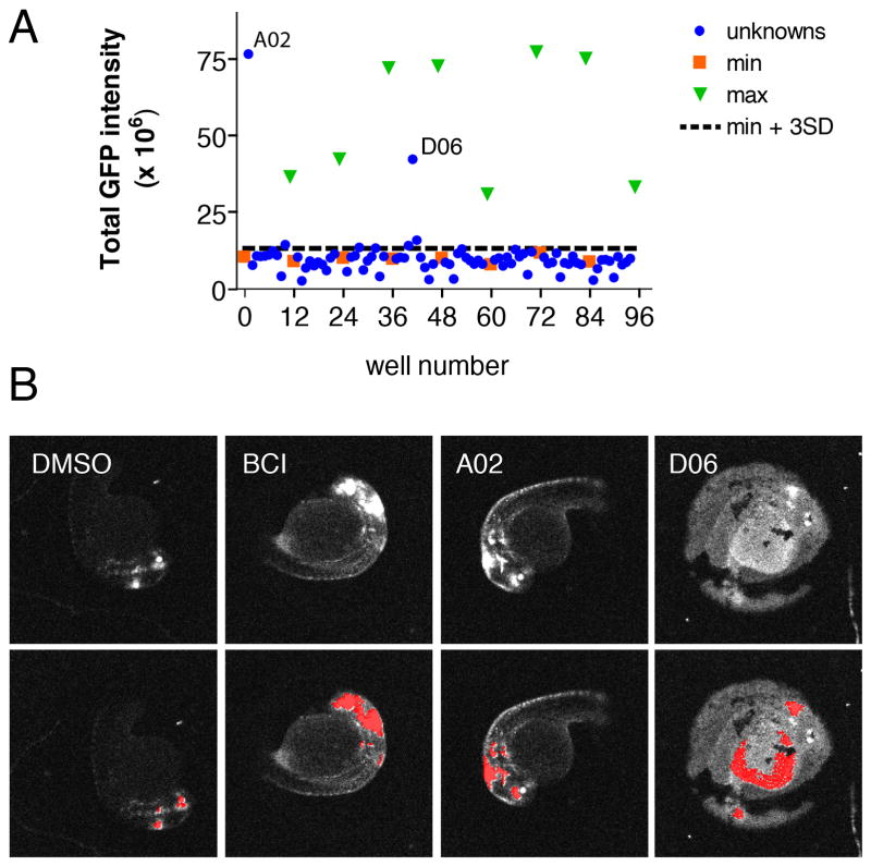
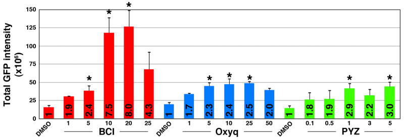
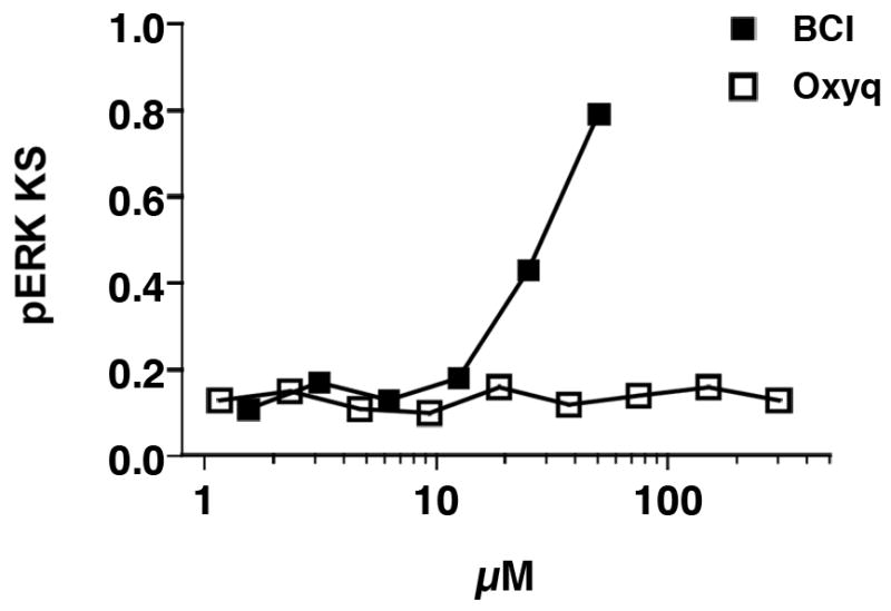
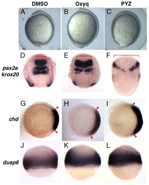
Zebrafish have become an invaluable vertebrate animal model to interrogate small molecule libraries for modulators of complex biological pathways and phenotypes. We have recently described the implementation of a quantitative, high-content imaging assay in multi-well plates to analyze the effects of small molecules on Fibroblast Growth Factor (FGF) signaling in vivo. Here we have evaluated the capability of the assay to identify compounds that hyperactivate FGF signaling from a test cassette of agents with known biological activities. Using a transgenic zebrafish reporter line for FGF activity, we screened 1040 compounds from an annotated library of known bioactive agents, including FDA-approved drugs. The assay identified two molecules, 8-hydroxyquinoline sulfate and pyrithione zinc, that enhanced FGF signaling in specific areas of the brain. Subsequent studies revealed that both compounds specifically expanded FGF target gene expression. Furthermore, treatment of early stage embryos with either compound resulted in dorsalized phenotypes characteristic of hyperactivation of FGF signaling in early development. Documented activities for both agents included activation of extracellular signal-related kinase (ERK), consistent with FGF hyperactivation. To conclude, we demonstrate the power of automated quantitative high-content imaging to identify small molecule modulators of FGF.


Figures

Fig 1
Fig 1
Automated chemical screen in zebrafish identifies Oxyq as a potential activator of FGF signaling. (A) Graph showing mean GFP intensity from two plates together with the mean + 3 SD limits of the negative control wells (B) Morphology of Tg(dusp6:d2eGFP) embryos in the presence of negative control (DMSO), positive control (BCI) and 8-hydroxyquinoline hemisulfate (A02). Well D06 illustrates the appearance of a fluorescent yolk that is not the site of FGF activity and this “hit” was eliminated. Upper panels show original scan images; lower panels show images with CNT analysis applied.
Fig 2
Fig 2
Oxyq and PYZ are mild activators of FGF signaling. A graphical representation of Tg(dusp6:d2eGFP) embryos after drug exposure. The total GFP intensity is expressed as normalized values and treatment response is represented as fold increase over the control (numbers in bars). BCI demonstrated an eight-fold increase whereas Oxyq and PYZ elicited two to three-fold increases in GFP intensity over the control. Treatment bars are represented as mean ± SE. Statistical significance was tested using Student’s t-test (two-tailed assuming unequal variances. *, p<0.05, n=8.
Fig 3
Fig 3
Oxyq is not a DUSP6 inhibitor in a cell based chemical complementation assay quantifying pERK levels in DUSP expressing cells. HeLa cells were transfected with c-Myc tagged DUSP6. Cells with high levels of c-Myc-tagged DUSP6 were identified based on c-Myc staining. pERK levels were measured in the DUSP expressing cells and a cumulative distribution function (cdf) assembled for each well. Cdfs of each well were compared to a reference distribution from 16 vehicle treated cells. Large KS values denote high dissimilarity from control and indicate high pERK levels in the DUSP expressing cells due to phosphatase inhibition. Data are the mean ± SE of four replicates from a single experiment that was repeated once with identical results.
Fig 4
Fig 4
Oxyq and PYZ treatment induced dorsalization in zebrafish. Morphology of zebrafish embryos treated with Oxyq and PYZ from 1K cells until early somitogenesis stage. The neural markers pax2a and krox20 showed lateral expansion (compare 4D to 4E and F). chd expression was expanded in treated groups over control (compare 4G to 4H and I). dusp6 expression showed mild expansion in OxyQ and PYZ treated groups over control.

References

    1. Chen HL, Chang CY, Lee HT, Lin HH, Lu PJ, Yang CN, Shiau CW, Shaw AY. Synthesis and pharmacological exploitation of clioquinol-derived copper-binding apoptosis inducers triggering reactive oxygen species generation and MAPK pathway activation. Bioorg Med Chem. 2009;17(20):7239–7247. - PubMed
    1. Collery RF, Link BA. Dynamic smad-mediated BMP signaling revealed through transgenic zebrafish. Dev Dyn. 2011;240(3):712–722. - PMC - PubMed
    1. de Groh ED, Swanhart LM, Cosentino CC, Jackson RL, Dai W, Kitchens CA, Day BW, Smithgall TE, Hukriede NA. Inhibition of histone deacetylase expands the renal progenitor cell population. J Am Soc Nephrol. 2010;21(5):794–802. - PMC - PubMed
    1. den Hertog J. Chemical genetics: Drug screens in Zebrafish. Biosci Rep. 2005;25(5–6):289–297. - PubMed
    1. Furthauer M, Van Celst J, Thisse C, Thisse B. Fgf signalling controls the dorsoventral patterning of the zebrafish embryo. Development. 2004;131(12):2853–2864. - PubMed

Publication types

MeSH terms

Substances

LinkOut - more resources