Comparative Study
. 2011 Oct 25;50(42):9066-75.
doi: 10.1021/bi201094v. Epub 2011 Sep 27.

Substitution of the human αC region with the analogous chicken domain generates a fibrinogen with severely impaired lateral aggregation: fibrin monomers assemble into protofibrils but protofibrils do not assemble into fibers

Affiliations
Comparative Study

Substitution of the human αC region with the analogous chicken domain generates a fibrinogen with severely impaired lateral aggregation: fibrin monomers assemble into protofibrils but protofibrils do not assemble into fibers

Lifang Ping et al. Biochemistry. .

Abstract

Fibrin polymerization occurs in two steps: the assembly of fibrin monomers into protofibrils and the lateral aggregation of protofibrils into fibers. Here we describe a novel fibrinogen that apparently impairs only lateral aggregation. This variant is a hybrid, where the human αC region has been replaced with the homologous chicken region. Several experiments indicate this hybrid human-chicken (HC) fibrinogen has an overall structure similar to normal. Thrombin-catalyzed fibrinopeptide release from HC fibrinogen was normal. Plasmin digests of HC fibrinogen produced fragments that were similar to normal D and E; further, as with normal fibrinogen, the knob 'A' peptide, GPRP, reversed the plasmin cleavage associated with addition of EDTA. Dynamic light scattering and turbidity studies with HC fibrinogen showed polymerization was not normal. Whereas early small increases in hydrodynamic radius and absorbance paralleled the increases seen during the assembly of normal protofibrils, HC fibrinogen showed no dramatic increase in scattering as observed with normal lateral aggregation. To determine whether HC and normal fibrinogen could form a copolymer, we examined mixtures of these. Polymerization of normal fibrinogen was markedly changed by HC fibrinogen, as expected for mixed polymers. When the mixture contained 0.45 μM normal and 0.15 μM HC fibrinogen, the initiation of lateral aggregation was delayed and the final fiber size was reduced relative to normal fibrinogen at 0.45 μM. Considered altogether, our data suggest that HC fibrin monomers can assemble into protofibrils or protofibril-like structures, but these either cannot assemble into fibers or assemble into very thin fibers.


Figures

Figure 1
Figure 1
Schematic of the junction of human and chicken Aα chain sequences. A segment of the human sequence (top line) is aligned with the similar segment of the chicken sequence (bottom line). Identical and similar residues are shaded. The human/chicken hybrid sequence includes human residues Ala1 through Arg197 joined to chicken residues Gln199 through Lys491. The encoded segment is shown in BOLD.
Figure 2
Figure 2
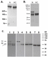
SDS-PAGE and immunoblot analysis of human/chicken fibrinogen. Fibrinogens were analyzed by SDS-PAGE run under non-reducing (8% polyacrylamide, A) and reducing (10% polyacrylamide, B) conditions, and stained with Coomassie blue. Immunoblots (C) were prepared from 10% gels run under reducing conditions. Blots were developed with: a polyclonal antibodies to human fibrinogen (lanes 1 and 2); a monoclonal antibody specific for the N-terminus of the Aα chain (Lanes 3 and 4); a monoclonal antibody specific for the N-terminus of the Bβ chain (lanes 5 and 6); and a monoclonal antibody specific for the C-terminus of the γ chain (lanes 7 and 8). For all figures, odd numbered lanes are normal fibrinogen and even numbered lanes are HC fibrinogen. Molecular weight markers are indicated at the left (A and B) and on the right (C).
Figure 3
Figure 3
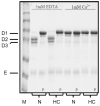
Plasmin digests. Normal (N) and human-chicken (HC) fibrinogens with and without peptide GPRP (P) were incubated with plasmin in the presence of 5 mM EDTA or 1 mM CaCl2. Samples were analyzed by SDS-PAGE (8%) run under non-reducing conditions. Normal plasmin digestion products D1, D2, D3 and E are indicated on the left.
Figure 4
Figure 4
Polymerization measured by turbidity (A) and dynamic light scattering (B). Polymerization was initiated by addition of thrombin to normal (solid line, solid squares) and HC (broken line, solid circles) fibrinogens. Representative turbidity curves (A) were obtained with 0.6 μM fibrinogen and 0.1 U/mL thrombin. The dotted line represents zero absorbance. Representative DLS curves (B) were obtained with 1.2 μM fibrinogen and 0.01 U/mL thrombin. The insert shows the same DLS data but on the different Y-scale.
Figure 5
Figure 5
Polymerization of fibrinogen mixtures. Polymerization was initiated by adding thrombin (0.1 U/mL) to normal or HC fibrinogens (0.6 μM) or their mixtures (mole/mole, 0.6 μM total fibrinogen). Polymer formation was measured by the change in turbidity with time. Representative curves (solid lines for all except the 50/50% N/HC mixture, broken line) from one experiment are shown. The dotted line represents zero absorbance.
Figure 6
Figure 6
Polymerization of fibrinogen monomers. Fibrinogen monomers were purified by gel-filtration chromatography. Representative results for thrombin-catalyzed (0.1 U/mL) polymerization of normal (N), HC and their mixtures (N/HC, percent of mole/mole). A representative polymerization experiment followed by DLS is shown in panels A and B. The presentation in panel B expands the Y-axis to show the time dependent change in radius for the relatively small forms, and extends the X-axis to show the gradually decreasing slope of the time-dependent change in radius for 100% HC (open circles) and the 50/50 normal/HC mixture (open triangles). A representative experiment followed by turbidity is shown in C and D. The presentation in panel D expands both axes to show the time dependent change at low turbidity at early times during polymerization. Both experiments were performed under conditions described in Figure 4.
Figure 7
Figure 7
Polymerization of the normal/HC mixture relative to normal fibrinogen. Polymerization of normal fibrinogen (solid lines) at 0.60 μM (100%), 0.45 μM (75%), and 0.30 μM (50%) and the 75:25 mixture (broken line) of 0.45 μM normal and 0.15 μM HC fibrinogens. Conditions were as described in Figure 4.
Figure 8
Figure 8
Schematic of the model for polymerization of the 25:75 mixture of normal and HC fibrinogens. Normal fibrinogen is represented by a blue rectangle, HC by a blue rectangle with a red drop. Initially, the normal and HC monomers (A) assemble stochastically into mixed protofibrils (B). These protofibrils are in equilibrium with the monomers. The assembly of fibers (C) is rapid and irreversible (fat arrow) for protofibrils that are able to form fibers and slow (thin arrow) for protofibrils that are incapable of lateral aggregation. Red stars represent clashes between protofibrils that prevent or impair lateral aggregation. Assuming that the protofibrils that can assemble into fibers have more normal monomers (i.e. less than 1 in 4 is HC monomer), the formation of fibers will lead to an increase in the representation of HC monomers in the monomer↔protofibril equilibrium. As the reaction proceeds, the assembly of fibers will approach that seen in the 50:50 mixture. Thus, we expect the final clot will be a mixture of fibers, ranging from those found with normal fibrinogen at 0.3 μM to those found with the 50:50 mixture.

References

    1. Kollman JM, Pandi L, Sawaya MR, Riley M, Doolittle RF. Crystal Structure of Human Fibrinogen. Biochemistry. 2009;48:3877–3886. - PubMed
    1. Medved L, Weisel JW. Recommendations for nomenclature on fibrinogen and fibrin. J Thromb Haemost. 2009;7:355–359. - PMC - PubMed
    1. Weisel JW, Medved L. The structure and function of the alpha C domains of fibrinogen. Ann N Y Acad Sci. 2001;936:312–327. - PubMed
    1. Medved LV, Gorkun OV, Privalov PL. Structural organization of C-terminal parts of fibrinogen A alpha-chains. FEBS Letters. 1983;160:291–295. - PubMed
    1. Tsurupa G, Hantgan RR, Burton RA, Pechik I, Tjandra N, Medved L. Structure, Stability, and Interaction of the Fibrin(ogen) αC-Domains. Biochemistry. 2009;48:12191–12201. - PMC - PubMed

Publication types

MeSH terms