. 2011 Sep 21;8(1):15.
doi: 10.1186/1559-0275-8-15.

Morphological changes in diabetic kidney are associated with increased O-GlcNAcylation of cytoskeletal proteins including α-actinin 4

Affiliations

Morphological changes in diabetic kidney are associated with increased O-GlcNAcylation of cytoskeletal proteins including α-actinin 4

Yoshihiro Akimoto et al. Clin Proteomics. .

Abstract

Purpose: The objective of the present study is to identify proteins that change in the extent of the modification with O-linked N-acetylglucosamine (O-GlcNAcylation) in the kidney from diabetic model Goto-Kakizaki (GK) rats, and to discuss the relation between O-GlcNAcylation and the pathological condition in diabetes.

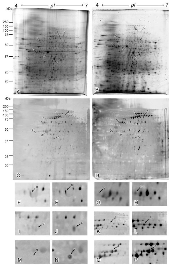
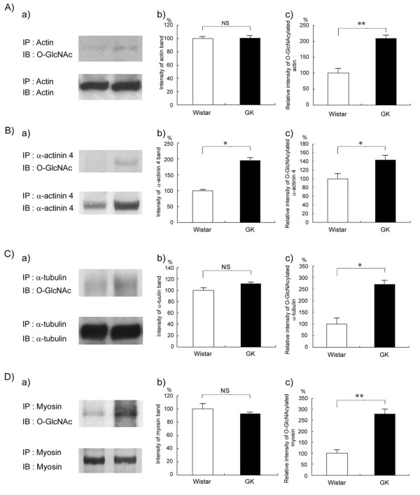
Methods: O-GlcNAcylated proteins were identified by two-dimensional gel electrophoresis, immunoblotting and peptide mass fingerprinting. The level of O-GlcNAcylation of these proteins was examined by immunoprecipitation, immunoblotting and in situ Proximity Ligation Assay (PLA).

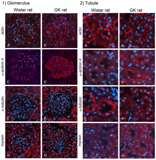
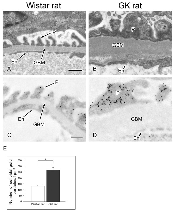
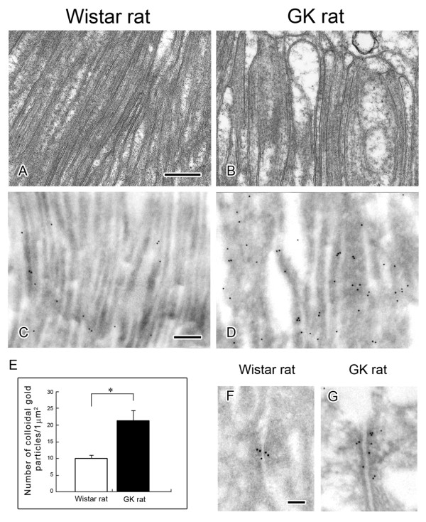
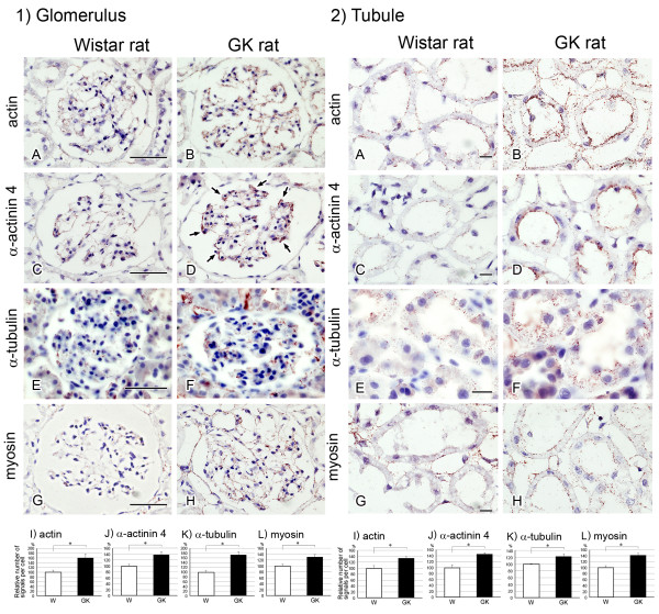
Results: O-GlcNAcylated proteins that changed significantly in the degree of O-GlcNAcylation were identified as cytoskeletal proteins (α-actin, α-tubulin, α-actinin 4, myosin) and mitochondrial proteins (ATP synthase β, pyruvate carboxylase). The extent of O-GlcNAcylation of the above proteins increased in the diabetic kidney. Immunofluorescence and in situ PLA studies revealed that the levels of O-GlcNAcylation of actin, α-actinin 4 and myosin were significantly increased in the glomerulus and the proximal tubule of the diabetic kidney. Immunoelectron microscopy revealed that immunolabeling of α-actinin 4 is disturbed and increased in the foot process of podocytes of glomerulus and in the microvilli of proximal tubules.

Conclusion: These results suggest that changes in the O-GlcNAcylation of cytoskeletal proteins are closely associated with the morphological changes in the podocyte foot processes in the glomerulus and in microvilli of proximal tubules in the diabetic kidney. This is the first report to show that α-actinin 4 is O-GlcNAcylated. α-Actinin 4 will be a good marker protein to examine the relation between O-GlcNAcylation and diabetic nephropathy.


Figures

Figure 1
Figure 1
Comparison of total protein map and O-GlcNAcylation map of kidney between nondiabetes and diabetes. (A, B) Representative total protein map of 2D PAGE for nondiabetic (A) and diabetic (B) rat kidneys detected by SYPRO-Ruby. (C, D) Typical O-GlcNAcylated protein map of 2D PAGE for nondiabetic (C) and diabetic (D) rat kidneys detected by O-GlcNAc antibody. Spots indicated by arrows represent the identified proteins that had changed in terms of O-GlcNAc level. (E-L) Regions around spots "a," "b," "c," "d," "e," and "f" in the maps "C" and "D" are enlarged to facilitate the identification of each spot, indicated by arrows, in the non-diabetic (E, G, I, K, M, O) and diabetic (F, H, J, L, N, P) kidneys.
Figure 2
Figure 2
Analysis of O-GlcNAcylation level of cytoskeletal proteins by immunoprecipitation and immunoblotting. Total lysates of nondiabetic and diabetic kidneys were immunoprecipitated with anti-actin (A), anti-α-actinin 4 (B), anti-α-tubulin antibody (C) or anti-myosin (D). The immunocomplexes were separated by SDS-PAGE, transferred to PVDF membranes, and probed with O-GlcNAc antibody (a, upper panel). The membrane was then stripped and reprobed with the antibody used for immunoprecipitation (a, lower panel). Results shown are representative of 3 independent experiments. Intensity of bands recognized by the antibody used for immunoprecipitation was quantified by scanning densitometry (b). Relative intensities of the O-GlcNAc reactive bands to bands reactive with each antibody used for immunoprecipitation were then determined (c). Data are the means ± SEM from 3 different rats. (□), Wistar rats; (■), GK rats. *P < 0.05 and **p < 0.01 vs. control Wistar rat.
Figure 3
Figure 3
Immunohistochemical analysis of cytoskeletal proteins by confocal laser scanning microscope. Localization of actin, α-actinin 4, myosin, and α-tubulin in glomeruli (1) and tubules (2). 1) Although the intensity of immunoreactivity of α-tubulin did not change, that of immunoreactivity of actin, α-actinin 4 and myosin was increased in the diabetic glomerulus. 2) Whereas the intensity of immunoreactivity of actin and myosin did not change, that of immunoreactive α-actinin 4 was increased and that of immunoreactive α-tubulin were decreased in the diabetic tubules. Scale bars, 1)-A, 50 μm, 2)-A, 20 μm.
Figure 4
Figure 4
Morphological change of foot processes of podocytes in glomerulus is correlated with the change of α-actinin 4. Electron microscopic appearance of glomerular capillary (A, B) and immuno-electron microscopic localization of α-actinin 4 in glomerular capillaries (C, D). (A, B) Comparison of electron micrographs of the capillary wall of the glomerulus from nondiabetic (A) and diabetic (B) kidneys revealed fused foot processes of podocytes and a thickened basement membrane in the sections from the diabetic rat. En, endothelial cell; GBM, glomerular basement membrane; P, foot process of podocyte. Scale bar, 200 nm. (C, D) Sections of nondiabetic and diabetic kidneys were immunolabeled for α-actinin 4 (12-nm gold particles). Whereas the gold particles were localized mainly in the periphery of the foot processes in the nondiabetic kidney, they were found not only in the periphery but also in the inner aspect of foot processes in the diabetic kidney. (E) Density (number/μm2) of colloidal gold particles representing α-actinin 4 in the foot processes of podocytes from nondiabetic (open bar) and diabetic (closed bar) kidneys. Data are the means ± SEM from 3 different rats. *P < 0.01 vs. control (Wistar rat).
Figure 5
Figure 5
Morphological change of microvilli in proximal tubule is correlated with the change of α-actinin 4. Electron microscopic appearance of microvilli of the tubules (A, B) and immuno-electron microscopic localization of α-actinin 4 in microvilli (C, D) and in the adherins junction (F, G) of tubules. (A, B) Comparison of electron micrographs of proximal tubules from nondiabetic rat (A) and diabetic rat (B) revealed that microvilli of the diabetic rat were swollen and had become bulbous, whereas those of the Wistar rat were regularly shaped. (C, D, F, G) Sections of nondiabetic and diabetic kidneys were immunolabeled for α-actinin 4 (12-nm gold particles). Whereas only a few gold particles were localized in the microvilli of proximal tubules in the nondiabetic kidney, many gold particles were detected in the microvilli of proximal tubules in the diabetic kidney (C, D). (E) Density (number/μm2) of colloidal gold particles representing α-actinin 4 in the microvilli from nondiabetic (open bar) diabetic kidney (closed bar) kidneys. Data are the means ± SEM from 3 different rats. *P < 0.01 vs. control (Wistar rat). (F, G) The colloidal gold labels indicating α-actinin 4 were also observed in the adherins junction of proximal tubules of both nondiabetic (F) and diabetic (G) kidneys. Scale bars: 200 nm (A, C) and 100 nm (F).
Figure 6
Figure 6
Analysis of O-GlcNAcylated cytoskeletal proteins by using the in situ PLA assay. Localization of O-GlcNAcylated actin (A, B), O-GlcNAcylated α-actinin 4 (C, D), O-GlcNAcylated α-tubulin (E, F) and O-GlcNAcylated myosin (G, H) in the glomerulus (1) and tubule (2) of normal (A, C, E, G) and diabetic (B, D, F, H) rats. Arrows in D indicate podocytes. I-L) Relative number of signals per cell. Ten different glomeruli and tubules were obtained from each sample. Signals were analyzed by Blob-Finder software. Values represent means ± SEM from 3 different rats. *P < 0.05 vs. control Wistar rat (W).

References

    1. Wells L, Vosseller K, Hart GW. Glycosylation of nucleocytoplasmic proteins: signal transduction and O-GlcNAc. Science. 2001;291:2376–2378. doi: 10.1126/science.1058714. - DOI - PubMed
    1. Hart GW, Housley MP, Slawson C. Cycling of O-linked β-N-acetylglucosamine on nucleocytoplasmic proteins. Nature. 2007;446:1017–1022. doi: 10.1038/nature05815. - DOI - PubMed
    1. Butkinaree C, Park K, Hart GW. O-linked β-N-acetylglucosamine (O-GlcNAc): Extensive crosstalk with phosphorylation to regulate signaling and transcription in response to nutrients and stress. Biochim Biophys Acta. 2010;1800:96–106. - PMC - PubMed
    1. Yang X, Ongusaha PP, Miles PD. et al. Phosphoinositide signaling links O-GlcNAc transferase to insulin resistance. Nature. 2008;451:964–969. doi: 10.1038/nature06668. - DOI - PubMed
    1. Teo CF, Wollaston-Hayden EE, Wells L. Hexosamine flux, the O-GlcNAc modification, and the development of insulin resistance in adipocytes. Mol Cell Endocrinol. 2010;318:44–53. doi: 10.1016/j.mce.2009.09.022. - DOI - PMC - PubMed

LinkOut - more resources