. 2011 Nov;179(5):2490-500.
doi: 10.1016/j.ajpath.2011.07.034. Epub 2011 Sep 18.

Collagen type II and a thermo-responsive polymer of N-isopropylacrylamide induce arthritis independent of Toll-like receptors: a strong influence by major histocompatibility complex class II and Ncf1 genes

Affiliations

Collagen type II and a thermo-responsive polymer of N-isopropylacrylamide induce arthritis independent of Toll-like receptors: a strong influence by major histocompatibility complex class II and Ncf1 genes

Akhilesh Kumar Shakya et al. Am J Pathol. 2011 Nov.

Abstract

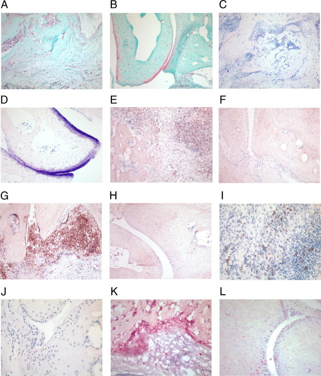
We established and characterized an arthritis mouse model using collagen type II (CII) and a thermo-responsive polymer, poly(N-isopropylacrylamide) (PNiPAAm). The new PNiPAAm adjuvant is TLR-independent, as all immunized TLR including MyD88-deficient mice developed an anti-CII response. Unlike other adjuvants, PNiPPAm did not skew the cytokine response (IL-1β, IFN-γ, IL-4, and IL-17), as there was no immune deviation towards any one type of immune spectrum after immunization with CII/PNiPPAm. Hence, using PNiPAAm, we studied the actual immune response to the self-protein, CII. We observed arthritis and autoimmunity development in several murine strains having different major histocompatibility complex (MHC) haplotypes after CII/PNiPAAm immunization but with a clear MHC association pattern. Interestingly, C57Bl/6 mice did not develop CII-induced arthritis, with PNiPAAm demonstrating absolute requirement for a classical adjuvant. Presence of a gene (Ncf1) mutation in the NADPH oxidation complex has a profound influence in arthritis and using PNiPAAm we could show that the high CIA severity in Ncf1 mutated mice is independent of any classical adjuvant. Macrophages, neutrophils, eosinophils, and osteoclasts but not mast cells dominated the inflamed joints. Furthermore, arthritis induction in the adjuvant-free, eosinophil-dependent Vβ12 DBA/1 mice could be shown to develop arthritis independent of eosinophils using CII/PNiPAAm. Thus, biocompatible and biodegradable PNiPAAm offers unique opportunities to study actual autoimmunity independent of TLR and a particular cytokine phenotype profile.


Figures

Figure 1
Figure 1
PNiPAAm-enhanced serum cytokine production. Groups of (BALB/c × B10.Q) F1 mice (n = 15) were immunized with 100 μg of rat CII emulsified with complete Freund's adjuvant (CFA-CII) or incomplete Freund's adjuvant (IFA-CII) or mixed with PNiPAAm. Sera collected on days 10 and 19 were analyzed for various cytokines, IFN-γ (A), IL-4 (B), IL-17 (C), and IL-1β (D) as described in Materials and Methods. CII without any adjuvant induced low level of IL-17 (0–2.2 ng/mL) but no detectable level of other cytokines (IFN-γ, IL-4, and IL-1β). Similarly, TNF-α levels were undetectable in all of the groups during this early phase of CIA (days 10 and 19). *P < 0.05; ***P < 0.001. Error bars denote ± SEM; n indicates number of mice in each group.
Figure 2
Figure 2
Influence of TLRs on PNiPAAm-CII–induced arthritis. Groups of TLR4 deleted (n = 8) and WT (n = 10) male mice on B10.Q genetic background were immunized with 100 μg of PNiPAAm-CII on day 0 and boosted on day 35 with 50 μg of PNiPAAm-CII. Arthritis incidence was 88% in TLR4 deficient and 20% in wild-type littermate controls. Female mice (7–11 mice per group) did not show any significant arthritis development (data not shown). Mean clinical score of arthritis severity (A) and anti-CII IgG serum levels (B) are shown. A group of different toll-like receptor (TLR)–deficient mice on C57Bl/6 genetic background were injected with 100 μg of PNiPAAm-CII on day 0 and boosted on day 21 with 50 μg of PNiPAAm-CII. Serum samples collected at day 40 were used to measure anti-CII antibody levels (C). PNiPAAm alone injected mice showed negligible level of anti-CII antibody response (data not shown). Mice deficient in TLR1 (n = 9), TLR2 (n = 10), TLR3 (n = 16), TLR5 (n = 9), TLR6 (n = 10), TLR7 (n = 15), TLR9 (n = 10), or the adaptor molecule MyD88 (n = 8) were used in this experiment. Affinity purified anti-CII IgG antibodies were used as standard. Error bars indicate ± SEM.
Figure 3
Figure 3
Genetic influence on PNiPAAm-CII susceptibility. Groups of mice from several different strains were injected with 100 μg of PNiPAAm-CII on day 0 and boosted on day 35 with 50 μg of PNiPAAm-CII. Incidence (A) of arthritis (percentage of affected mice) and arthritis severity (B) are shown. B10.Q (H-2q; n = 25), DBA/1 (H-2q; n = 26), C3H.Q (H-2q; n = 13), QD (H-2q; n = 18), QB (H-2q/d; n = 19), B10.P (H-2p; n = 25), C57Bl/6 (H-2b; n = 22), and BALB/c (H-2d; n = 15) mice were used in this experiment. Serum anti-CII IgG levels (C) from several different mouse strains at days 35 and 70. Mice with MHC haplotype (H-2q) were highly susceptible (cumulative incidence of arthritis in H-2q versus other MHC haplotypes, P < 0.0001) with a more severe arthritis phenotype (mean maximal score, P < 0.0001) and a robust adaptive immune response (C) whereas other MHC haplotypes (H-2p, b, d) were arthritis resistant with low anti-CII antibody levels (day 35, P < 0.05 and day 70, P < 0.0001). Injecting polymer alone or with ovalbumin did not induce arthritis in the highly arthritis-prone QD mice. Pooled sera from PNiPAAm-CII mice were used as standard. Error bar indicates ± SEM; n indicates the number of mice in each group. *P < 0.05 and ****P < 0.0001.
Figure 4
Figure 4
Presence of a low oxidative environment significantly enhanced PNiPAAm-CII arthritis. Groups of Ncf1 mutated (Ncf1*/*; n = 17) and wild-type (WT; n = 16) littermate controls were immunized at the base of the tail with 100 μg of PNiPAAm-CII on day 0 and boosted on day 35 with 50 μg of PNiPAAm-CII. Incidence (A) of arthritis (percentage of affected mice), mean clinical score of arthritis severity (B), and serum anti-CII IgG (C) and IgG subclass (D) levels in Ncf1*/* and WT mice at days 35 and 70 are shown. Affinity purified anti-CII IgG antibodies and pooled sera from PNiPAAm-CII mice were used as standards. Error bars indicate ± SEM; n denotes number of mice in each group. *P < 0.05 and ****P < 0.0001.
Figure 5
Figure 5
Histology of paws from PNiPAAm-CII–immunized Ncf1 and wild-type littermate control mice. Paws were taken from PNiPAAm-CII–immunized mice on day 62. Safranin staining for joint morphology and mast cells (A and B), and toluidine staining for proteoglycan depletion (C and D) from Ncf1 and WT mice. Stained for RB6+ cells (E and F), Mac1+ cells (G and H), cyanide-resistant eosinophil peroxidase activity (I and J), and tartarate-resistant acid phosphatase activity (K and L) in Ncf1 and WT mouse paws. Surrounding tissues of the joints showing the nature of infiltrating immune cells in the inflamed paw. Results shown are representative of those obtained from three to four mice in each group. Original magnifications, ×20.
Figure 6
Figure 6
CII-specific T cells significantly enhanced PNiPAAm-CII arthritis. Groups of Vβ12 transgenic (Vβ12 tg; n = 12) and nontransgenic littermate controls (WT; n = 21) littermate controls were immunized at the base of the tail with 100 μg of PNiPAAm-CII on day 0 and boosted on day 35 with 50 μg of PNiPAAm-CII. Incidence (A) of arthritis (percentage of affected mice), mean clinical score of arthritis severity (B), serum anti-CII IgG (C) levels in Vβ12 transgenic and nontransgenic littermate controls at days 35 and 70 are shown. Affinity purified anti-CII IgG antibodies were used as standard. IL-5 was blocked in vivo to address its role in PNiPAAm-CII–induced arthritis. Anti–IL-5 treatment in Vβ12tg mice did not have any significant role in arthritis incidence (D) or severity (E). Vβ12tg DBA/1 mice (n = 9 per group) was injected three times with a mixture of the IL-5–blocking antibody 50 μg of TRFK-5 mixed with 50 μg of affinity purified normal rat serum IgG or with the equivalent amount of pure normal rat IgG as control at days −2, 5, and 18. At day 0, all mice were immunized with 100 μg of PNiPAAm-CII at the base of the tail. Mice were clinically scored for arthritis until day 32. Error bars indicate ± SEM; n denotes number of mice in each group. *P < 0.05, **P < 0.01, and ***P < 0.001.

References

    1. Brunsberg U., Gustafsson K., Jansson L., Michaëlsson E., &Ährlund-Richter L., Pettersson S., Mattsson R., Holmdahl R. Expression of a transgenic class II Ab gene confers susceptibility to collagen-induced arthritis. Eur J Immunol. 1994;24:1698–1702. - PubMed
    1. Rosloniec E.F., Brand D.D., Myers L.K., Whittington K.B., Gumanovskaya M., Zaller D.M., Woods A., Altmann D.M., Stuart J.M., Kang A.H. An HLA-DR1 transgene confers susceptibility to collagen-induced arthritis elicited with human type II collagen. J Exp Med. 1997;185:1113–1122. - PMC - PubMed
    1. Wooley P.H., Luthra H.S., Stuart J.M., David C.S. Type II collagen-induced arthritis in mice: I. Major histocompatibility complex (I region) linkage and antibody correlates. J Exp Med. 1981;154:688–700. - PMC - PubMed
    1. Wooley P.H., Luthra H.S., Griffiths M.M., Stuart J.M., Huse A., David C.S. Type II collagen induced arthritis in mice: IV. Variations in immunogenetic regulation provide evidence for multiple arthritogenic epitopes on the collagen molecule. J Immunol. 1985;135:2443–2451. - PubMed
    1. Campbell I.K., Hamilton J.A., Wicks I.P. Collagen-induced arthritis in C57BL/6 (H-2b) mice: new insights into an important disease model of rheumatoid arthritis. Eur J Immunol. 2000;30:1568–1575. - PubMed

Publication types

MeSH terms