. 2012 Feb 1;59(3):2430-7.
doi: 10.1016/j.neuroimage.2011.09.003. Epub 2011 Sep 10.

NIRS-based hyperscanning reveals increased interpersonal coherence in superior frontal cortex during cooperation

Affiliations

NIRS-based hyperscanning reveals increased interpersonal coherence in superior frontal cortex during cooperation

Xu Cui et al. Neuroimage. .

Abstract

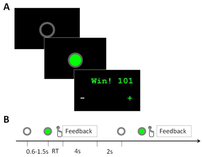
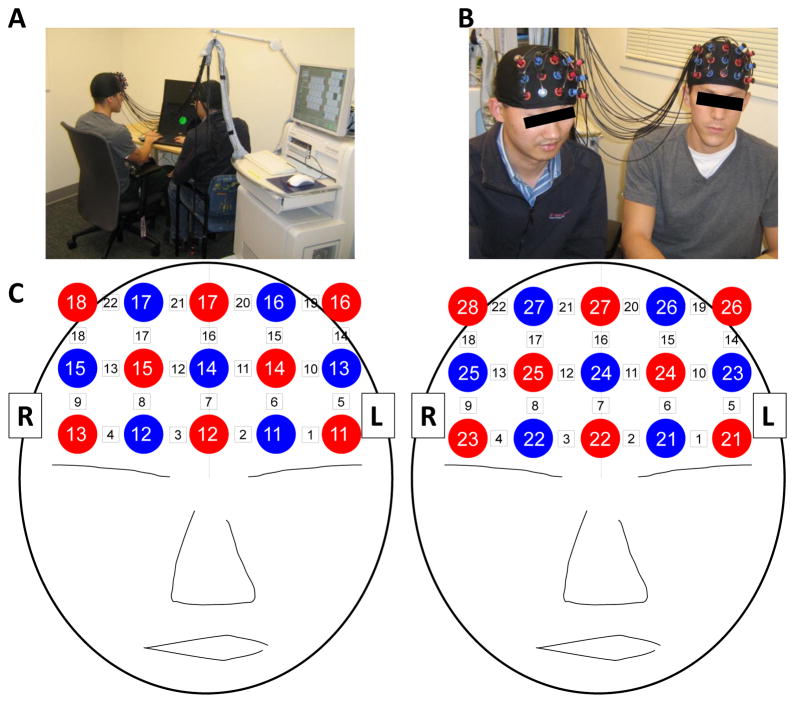
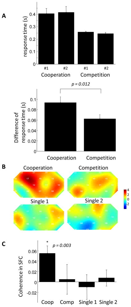
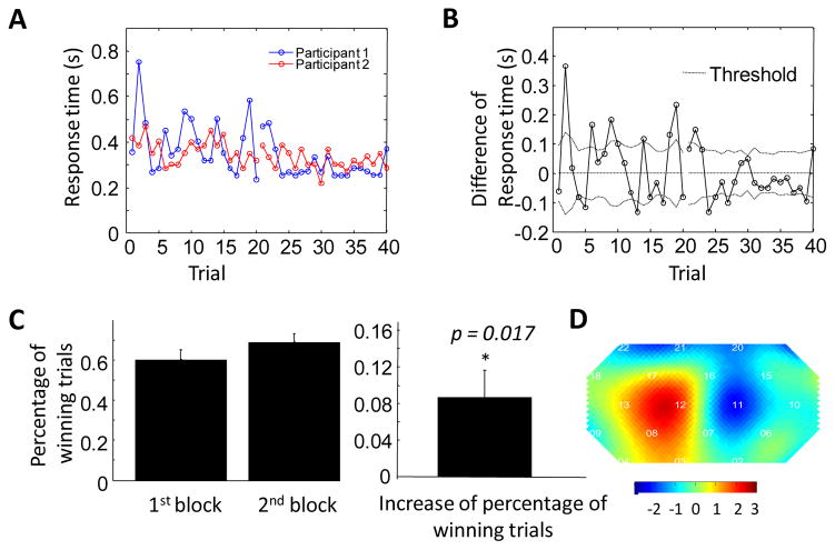
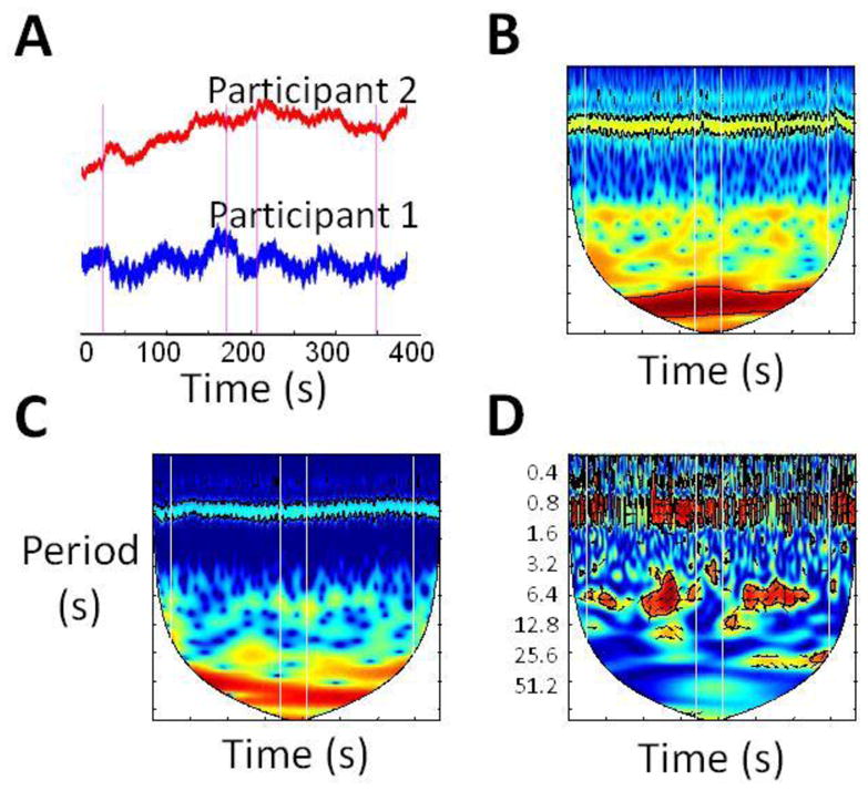
We used Near-Infrared Spectroscopy (NIRS) to simultaneously measure brain activity in two people while they played a computer-based cooperation game side by side. Inter-brain activity coherence was calculated between the two participants. We found that the coherence between signals generated by participants' right superior frontal cortices increased during cooperation, but not during competition. Increased coherence was also associated with better cooperation performance. To our knowledge, this work represents the first use of a single NIRS instrument for simultaneous measurements of brain activity in two people. This study demonstrates the use of NIRS-based hyperscanning in studies of social interaction in a naturalistic environment.


Conflict of interest statement

Competing Interest

The authors assert that they have no competing interests.

Figures

Figure 1
Figure 1
Task flow of the cooperation experiment. (A) Screenshots of the ready signal, “go” signal, and feedback window in a single trial. (B) Time flow of the experiment, showing two consecutive trials. The entire cooperation task consisted of two task blocks separated by a 30s rest period. Each task block consisted of 20 trials. RT: Response time.
Figure 2
Figure 2
Experimental Setup. (A & B) A pair of participants demonstrate the experimental setup. (C) Cap Configuration. Red circles indicate emitters; blue circles indicate detectors. White squares indicate measurement channels between emitters and detectors.
Figure 3
Figure 3
The inter-brain coherence increases in the superior frontal cortex during cooperation. (A) Wavelet transform coherence (WTC) between the raw oxy-Hb signal from channel 17 from the 1st participant and the raw oxy-Hb signal from channel 17 from the 2nd participant in a representative participant pair. The vertical white lines indicate the onset and offset timing of the task block. The coherence, encoded by color, is higher during task than during rest in the task frequency band (3.2–12.8s). The high coherence in the ~0.8s frequency band is attributable to participant heartbeat. (B) Wavelet coherence in all channels in participant pair #1. The relative position of channels is identical to that shown in Figure 2, where the upper rows cover the superior frontal cortex while the lower rows cover the inferior frontal cortex. It is seen that the coherence increases during the task in the superior frontal cortex. (C) Group analysis of coherence increase in all 11 pairs of participants. Color indicates the t value. Coherence increase in channels 17 and 12, which are located in the right superior frontal cortex, is significant (p<0.05, FDR corrected).
Figure 4
Figure 4
Coherence increase is not significant during the competition, single 1 and single 2 experiments. (A) The response time (top) and the difference of response time between the two participants (bottom) are plotted against the task type. The difference between response times of the two participants is smaller during the competition task than during the cooperation task. (B) The group t-test map of coherence increase in the four tasks. The coherence increase is significant only in the cooperation task. (C) The amplitude of coherence in superior frontal cortex (channel 12 and 17) in the four tasks.
Figure 5
Figure 5
The inter-brain coherence is higher during the 2nd task block in the cooperation experiment. (A) Response times of the two participants in a representative pair as a function of trial number. It can be seen that the response time is closer in the 2nd block. (B) The difference between response times as a function of trial number. (C) The percentage of winning trials in the first and second cooperation block (left), and the increase in percentage of winning trials between the first and second cooperation task blocks, averaged across all participants. The percentage of winning trials in the second block is significantly higher than that in the first block, demonstrating a learning process. (D) t-test map of coherence change (coherence in the 2nd block minus that in the 1st block) shows that coherence in right frontal cortex is significantly higher in the second cooperation task block (p<0.02).
Figure 6
Figure 6
Hyperscanning reveals patterns not detectable by single-person brain scanning. (A) Raw oxy-Hb time courses from corresponding channels in each participant in a representative pair (i.e. channel 17 in each participant). Vertical lines indicate cooperation task onsets and offsets alternatingly. A 30s rest block separates the two task blocks. There is no obvious change in signal amplitude during the task block. (B & C) Continuous wavelet transform of time series from participant 1 and 2, respectively. There is no obvious change of power in the frequency domain during the task blocks. (D) Wavelet coherence between time courses from participants 1 and 2. The relationship between time courses is shown by high coherence at task-specific frequency.

References

    1. Adolphs R. Cognitive neuroscience of human social behaviour. Nat Rev Neurosci. 2003;4:165–78. - PubMed
    1. Astolfi L, Toppi J, De Vico Fallani F, Vecchiato G, Salinari S, Mattia D, Cincotti F, Babiloni F. Neuroelectrical hyperscanning measures simultaneous brain activity in humans. Brain Topogr. 2010;23:243–56. - PubMed
    1. Babiloni F, Cincotti F, Mattia D, Mattiocco M, De Vico Fallani F, Tocci A, Bianchi L, Marciani MG, Astolfi L. Hypermethods for EEG hyperscanning. Conf Proc IEEE Eng Med Biol Soc. 2006;1:3666–9. - PubMed
    1. Boecker M, Buecheler MM, Schroeter ML, Gauggel S. Prefrontal brain activation during stop-signal response inhibition: an event-related functional near-infrared spectroscopy study. Behavioural brain research. 2007;176:259–66. - PubMed
    1. Chang C, Glover GH. Time-frequency dynamics of resting-state brain connectivity measured with fMRI. Neuroimage. 2010;50:81–98. - PMC - PubMed

Publication types