. 2011 Nov;152(11):4298-309.
doi: 10.1210/en.2011-1521. Epub 2011 Sep 20.

Molecular properties of Kiss1 neurons in the arcuate nucleus of the mouse

Affiliations

Molecular properties of Kiss1 neurons in the arcuate nucleus of the mouse

Michelle L Gottsch et al. Endocrinology. 2011 Nov.

Abstract

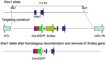
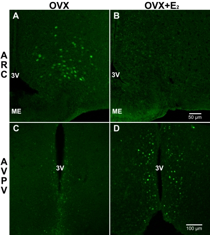
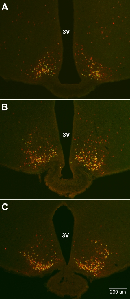
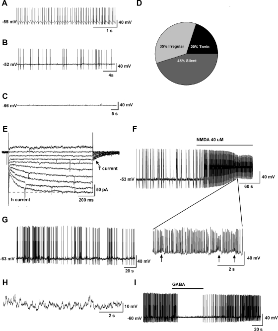
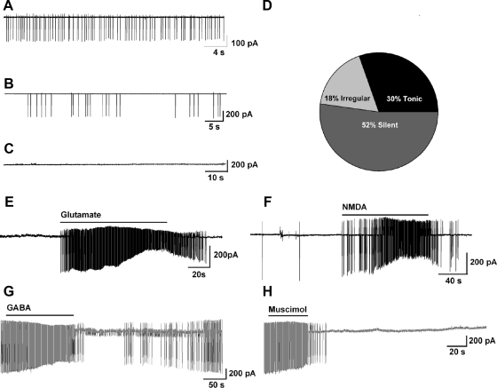
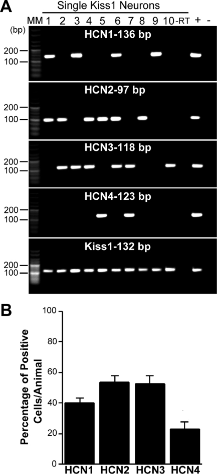
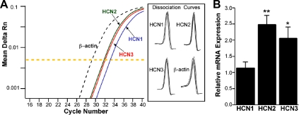
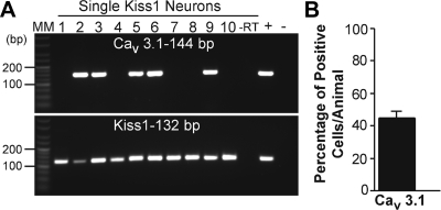
Neurons that produce kisspeptin play a critical role in reproduction. However, understanding the molecular physiology of kisspeptin neurons has been limited by the lack of an in vivo marker for those cells. Here, we report the development of a Kiss1-CreGFP knockin mouse, wherein the endogenous Kiss1 promoter directs the expression of a Cre recombinase-enhanced green fluorescent protein (GFP) fusion protein. The pattern of GFP expression in the brain of the knockin recapitulates what has been described earlier for Kiss1 in the male and female mouse, with prominent expression in the arcuate nucleus (ARC) (in both sexes) and the anteroventral periventricular nucleus (in females). Single-cell RT-PCR showed that the Kiss1 transcript is expressed in 100% of GFP-labeled cells, and the CreGFP transcript was regulated by estradiol in the same manner as the Kiss1 gene (i.e. inhibited in the ARC and induced in the anteroventral periventricular nucleus). We used this mouse to evaluate the biophysical properties of kisspeptin (Kiss1) neurons in the ARC of the female mouse. GFP-expressing Kiss1 neurons were identified in hypothalamic slice preparations of the ARC and patch clamped. Whole-cell (and loose attached) recordings revealed that Kiss1 neurons exhibit spontaneous activity and expressed both h- (pacemaker) and T-type calcium currents, and hyperpolarization-activated cyclic nucleotide-regulated 1-4 and CaV3.1 channel subtypes (measured by single cell RT-PCR), respectively. N-methyl-D-aspartate induced bursting activity, characterized by depolarizing/hyperpolarizing oscillations. Therefore, Kiss1 neurons in the ARC share molecular and electrophysiological properties of other CNS pacemaker neurons.


Figures

Fig. 1.
Fig. 1.
Kiss1-CreGFP targeting construct. A 13.2-kb Afe1-Sal1 fragment, including approximately 5.0-kb DNA 5′of the Kiss1 translation start site and approximately 8 kb on the 3′ side (depicted as broken dark blue arrow with a line through it in the top panel), was subcloned from a C57Bl/6 BAC clone into Bluescript. The targeting construct (depicted in the middle panel) contained a CreGFP fusion protein cassette (red and green arrow inserted in the middle of the first segment of the dark blue broken arrow from the top panel), followed by an frt-flanked SvNeo gene [for positive selection, illustrated by red diamonds (frt sites) surrounding a green arrow (SvNeo)] inserted just upstream of the normal start site for Kiss1. The construct also contained Pgk-DTA and HSV-TK (light blue arrows) genes for negative selection. The frt-Neo gene was removed by breeding the heterozygous mice with FLPer mice. The final construct is depicted in the bottom panel, illustrating a new Kiss1 allele that contains a Cre-eGFP cassette inserted before the native Kiss1 start site.
Fig. 2.
Fig. 2.
Photomicrographs of GFP-expressing cells in the ARC and the AVPV of OVX Kiss1-CreGFP knockin mice with and without E2 treatment. The distributions of GFP expression in the ARC (A and B) and the AVPV (C and D) are similar to the known distributions of Kiss1 mRNA in those same regions. Furthermore, E2 treatment reduces GFP expression in the ARC (B) compared with controls (A) but increases GFP expression in the AVPV (D) compared with controls (C), just as E2 has been shown to inhibit Kiss1 mRNA expression in the ARC and induce its expression in the AVPV (5). A and B show endogenous GFP fluorescence. In C and D, fluorescent immunohistochemistry was used to improve the visualization of GFP. ME, Median eminence; 3V, third ventricle.
Fig. 3.
Fig. 3.
Photomicrographs of GFP (green)- and β-Gal (red)-stained neurons in the ARC of OVX female Kiss1-CreGFP:LacZRep mice. Neurons labeled for both GFP and β-Gal appear yellow. Panels show sections from the rostral (A), middle (B), and caudal (C) arcuate. 3V, Third ventricle.
Fig. 4.
Fig. 4.
Electrophysiological characteristics of arcuate Kiss1 neurons in oil-treated OVX Kiss1-CreGFP mice using whole-cell patch recording. Kiss1 neurons in the ARC (of the female) rest at −63.8 ± 2.3 mV (n = 20). A–C, Representative traces of action potentials recorded from arcuate Kiss1 neurons showing tonic (A), irregular (B), and silent (C) firing patterns. D, Summary pie chart of the firing pattern distribution in ARC Kiss1 neurons (n = 20). E–H, Endogenous conductances and NMDA-induced burst firing of an ARC Kiss1 neuron. E, Ensemble of currents in response to depolarizing/hyperpolarizing steps from −50 to −140 mV illustrating the expression of a hyperpolarization-activated cation current (h-current) and a T-type Ca2+ current (arrow) in a representative Kiss1 neuron. Vhold = −60 mV. F, Current clamp recording in an ARC Kiss1 neuron showing the response to NMDA (40 μm). The spiking in F was expanded to illustrate the pronounced effects of NMDA on burst firing activity of Kiss1 neurons with an ensemble of spikes riding on top of low-threshold spikes (arrows). G, In a current clamped state close to the resting membrane potential, one can see the bursting activity in the presence of NMDA. H, In the presence of tetrodotoxin to block the fast Na+ spikes, one can clearly see the membrane oscillations (up and down states) induced by NMDA. Eighty percent of ARC Kiss1 neurons expressed the endogenous conductances (h-current, T-current) that are critical for generating burst firing activity. Although ARC Kiss1 neurons did not exhibit spontaneous burst firing activity, glutamate (NMDA) was able to induce burst firing activity in all of the cells. I, GABA (100 μm) inhibited firing in another arcuate kisspeptin neuron. Drugs were rapidly perfused into the bath as a 100-μl bolus.
Fig. 5.
Fig. 5.
Cellular characterization of arcuate Kiss1 neurons in oil-treated, OVX Kiss1-CreGFP mice in loose-patch cell recordings. A–C, Representative traces of action potentials recorded from Kiss1 neurons showing tonic (A), irregular (B), and silent (C) firing patterns. D, Summary pie chart of the firing pattern distribution in arcuate Kiss1 neurons (n = 23). E and F, Glutamate-induced (100 μm, n = 4) (E) and NMDA-induced (40 μm, n = 4) (F) burst firing in arcuate kisspeptin neurons. G and H, GABA-inhibited (100 μm, n = 5) (G) and muscimol-inhibited (10 μm, n = 4) (H) firing in arcuate Kis1 neurons. Drugs were rapidly perfused into the bath as a 100-μl bolus.
Fig. 6.
Fig. 6.
Expression of HCN channels in single Kiss1 neurons from the ARC of OVX Kiss1-CreGFP mice. A, Representative gels illustrating the mRNA expression of HCN channel subtypes 1–4. The expected sizes for the PCR products are as follows: for HCN1, 136 bp; for HCN2, 97 bp; for HCN3, 118 bp; for HCN4; 123 bp; and for Kiss1, 132 bp. As a negative control, a cell reacted −RT did not express any of the transcripts. MBH tissue RNA was also included as a positive control (+, With RT) and negative control (−, Without RT). MM, Molecular markers. B, Summary bar graphs of the percentage expression of HCN1, HCN2, HCN3, and HCN4. An average of 25 cells/animal from six animals was analyzed by scRT-PCR, and the mean number of neurons expressing HCN channel subtypes from each animal was determined. Bar graphs represent the mean ± sem of the percentage of Kiss1 neurons expressing each HCN subtype per animal.
Fig. 7.
Fig. 7.
Relative HCN subtype expression in Kiss1 neurons in the ARC of Kiss1-CreGFP mice. A, Real-time PCR amplification and dissociation curves for HCN1–3 and β-actin. The HCN1 (blue line), HCN2 (green line), and HCN3 (red line) depict the mean CT values for each of the three channel subtypes. The expression values were normalized to β-actin (black dotted line). The yellow dotted line is the midpoint of the exponential phase of amplification at which the CT was determined. B, qPCR measurements of HCN1, HCN2, and HCN3 mRNA in Kiss1 neuronal pools (three to six pools per animal) from OVX mice (n = 3 animals). HCN2 and HCN3 had higher expression relative to HCN1. The expression values were calculated via the ΔΔCT method and normalized to the mean ΔCT of HCN1 expression levels. Bar graphs represent the mean ± sem. *, P < 0.05, HCN3 vs. HCN1 (ANOVA); **, P < 0.01, HCN2 vs. HCN1 (ANOVA).
Fig. 8.
Fig. 8.
Expression the T-type Ca2+ channel CaV3.1 subtype in single Kiss1 neurons of female Kiss1-CreGFP OVX female mice. A, A representative gel illustrating the expression of CaV 3.1 and Kiss1 mRNA in Kiss1 neurons of the ARC. As a negative control, a cell reacted −RT did not express any of the transcripts. BH tissue was also included as a positive control (+, With RT) and negative control (−, Without RT). MM, Molecular markers. B, Summary bar graph of percentage expression of CaV3.1 in Kiss1 ARC neurons. An average of 20 cells/animal were harvested from six mice and were analyzed for CaV3.1 expression by scRT-PCR. Bar graphs represent the mean ± sem of the percentage of Kiss1 neurons expressing CaV3.1 per animal.

References

    1. de Roux N, Genin E, Carel JC, Matsuda F, Chaussain JL, Milgrom E. 2003. Hypogonadotropic hypogonadism due to loss of function of the KiSS1-derived peptide receptor GPR54. Proc Natl Acad Sci USA 100:10972–10976 - PMC - PubMed
    1. Seminara SB, Messager S, Chatzidaki EE, Thresher RR, Acierno JS, Jr, Shagoury JK, Bo-Abbas Y, Kuohung W, Schwinof KM, Hendrick AG, Zahn D, Dixon J, Kaiser UB, Slaugenhaupt SA, Gusella JF, O'Rahilly S, Carlton MB, Crowley WF, Jr, Aparicio SA, Colledge WH. 2003. The GPR54 gene as a regulator of puberty. N Engl J Med 349:1614–1627 - PubMed
    1. Gottsch ML, Cunningham MJ, Smith JT, Popa SM, Acohido BV, Crowley WF, Seminara S, Clifton DK, Steiner RA. 2004. A role for kisspeptins in the regulation of gonadotropin secretion in the mouse. Endocrinology 145:4073–4077 - PubMed
    1. Mikkelsen JD, Simonneaux V. 2009. The neuroanatomy of the kisspeptin system in the mammalian brain. Peptides 30:26–33 - PubMed
    1. Smith JT, Cunningham MJ, Rissman EF, Clifton DK, Steiner RA. 2005. Regulation of Kiss1 gene expression in the brain of the female mouse. Endocrinology 146:3686–3692 - PubMed

Publication types