Homocysteine promotes human endothelial cell dysfunction via site-specific epigenetic regulation of p66shc
- PMID: 21933910
- PMCID: PMC3211975
- DOI: 10.1093/cvr/cvr250
Homocysteine promotes human endothelial cell dysfunction via site-specific epigenetic regulation of p66shc
Abstract
Aims: Hyperhomocysteinaemia is an independent risk factor for atherosclerotic vascular disease and is associated with vascular endothelial dysfunction. Homocysteine modulates cellular methylation reactions. P66shc is a protein that promotes oxidative stress whose expression is governed by promoter methylation. We asked if homocysteine induces endothelial p66shc expression via hypomethylation of CpG dinucleotides in the p66shc promoter, and whether p66shc mediates homocysteine-stimulated endothelial cell dysfunction.
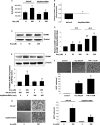
Methods and results: Homocysteine stimulates p66shc transcription in human endothelial cells and hypomethylates specific CpG dinucleotides in the human p66shc promoter. Knockdown of p66shc inhibits the increase in reactive oxygen species, and decrease in nitric oxide, elicited by homocysteine in endothelial cells and prevents homocysteine-induced up-regulation of endothelial intercellular adhesion molecule-1. In addition, knockdown of p66shc mitigates homocysteine-induced adhesion of monocytes to endothelial cells. Inhibition of DNA methyltransferase activity or knockdown of DNA methyltransferase 3b abrogates homocysteine-induced up-regulation of p66shc. Comparison of plasma homocysteine in humans with coronary artery disease shows a significant difference between those with highest and lowest p66shc promoter CpG methylation in peripheral blood leucocytes.
Conclusion: Homocysteine up-regulates human p66shc expression via hypomethylation of specific CpG dinucleotides in the p66shc promoter, and this mechanism is important in homocysteine-induced endothelial cell dysfunction.
Figures


Similar articles
-
Epigenetic upregulation of p66shc mediates low-density lipoprotein cholesterol-induced endothelial cell dysfunction.Am J Physiol Heart Circ Physiol. 2012 Jul 15;303(2):H189-96. doi: 10.1152/ajpheart.01218.2011. Epub 2012 Jun 1. Am J Physiol Heart Circ Physiol. 2012. PMID: 22661506 Free PMC article.
-
Canonical Wnt signaling induces vascular endothelial dysfunction via p66Shc-regulated reactive oxygen species.Arterioscler Thromb Vasc Biol. 2014 Oct;34(10):2301-9. doi: 10.1161/ATVBAHA.114.304338. Epub 2014 Aug 21. Arterioscler Thromb Vasc Biol. 2014. PMID: 25147340 Free PMC article.
-
Inhibition of S-Adenosylhomocysteine Hydrolase Induces Endothelial Dysfunction via Epigenetic Regulation of p66shc-Mediated Oxidative Stress Pathway.Circulation. 2019 May 7;139(19):2260-2277. doi: 10.1161/CIRCULATIONAHA.118.036336. Circulation. 2019. PMID: 30773021
-
Nitric oxide, oxidative stress, and p66Shc interplay in diabetic endothelial dysfunction.Biomed Res Int. 2014;2014:193095. doi: 10.1155/2014/193095. Epub 2014 Mar 5. Biomed Res Int. 2014. PMID: 24734227 Free PMC article. Review.
-
Apoptosis and oxidative stress-related diseases: the p66Shc connection.Curr Mol Med. 2009 Apr;9(3):392-8. doi: 10.2174/156652409787847254. Curr Mol Med. 2009. PMID: 19355920 Review.
Cited by
-
Ly6C+ Inflammatory Monocyte Differentiation Partially Mediates Hyperhomocysteinemia-Induced Vascular Dysfunction in Type 2 Diabetic db/db Mice.Arterioscler Thromb Vasc Biol. 2019 Oct;39(10):2097-2119. doi: 10.1161/ATVBAHA.119.313138. Epub 2019 Aug 1. Arterioscler Thromb Vasc Biol. 2019. PMID: 31366217 Free PMC article.
-
Homocysteine levels in patients with coronary slow flow phenomenon: A meta-analysis.PLoS One. 2023 Jul 7;18(7):e0288036. doi: 10.1371/journal.pone.0288036. eCollection 2023. PLoS One. 2023. PMID: 37418362 Free PMC article.
-
Homocysteine concentration and adenosine A2A receptor production by peripheral blood mononuclear cells in coronary artery disease patients.J Cell Mol Med. 2020 Aug;24(16):8942-8949. doi: 10.1111/jcmm.15527. Epub 2020 Jun 29. J Cell Mol Med. 2020. PMID: 32599677 Free PMC article.
-
Lipopolysaccharide Downregulates Kruppel-Like Factor 2 (KLF2) via Inducing DNMT1-Mediated Hypermethylation in Endothelial Cells.Inflammation. 2017 Oct;40(5):1589-1598. doi: 10.1007/s10753-017-0599-0. Inflammation. 2017. PMID: 28578476
-
Leveraging clinical epigenetics in heart failure with preserved ejection fraction: a call for individualized therapies.Eur Heart J. 2021 May 21;42(20):1940-1958. doi: 10.1093/eurheartj/ehab197. Eur Heart J. 2021. PMID: 33948637 Free PMC article. Review.
References
-
- Kang SS, Wong PW, Malinow MR. Hyperhomocyst(e)inemia as a risk factor for occlusive vascular disease. Annu Rev Nutr. 1992;12:279–298. - PubMed
-
- Stampfer MJ, Malinow MR, Willett WC, Newcomer LM, Upson B, Ullmann D, et al. A prospective study of plasma homocyst(e)ine and risk of myocardial infarction in US physicians. JAMA. 1992;268:877–881. - PubMed
-
- McCully KS. Homocysteine and vascular disease. Nat Med. 1996;2:386–389. - PubMed
-
- Bonaa KH, Njolstad I, Ueland PM, Schirmer H, Tverdal A, Steigen T, et al. Homocysteine lowering and cardiovascular events after acute myocardial infarction. N Engl J Med. 2006;354:1578–1588. - PubMed
-
- Lonn E, Yusuf S, Arnold MJ, Sheridan P, Pogue J, Micks M, et al. Homocysteine lowering with folic acid and B vitamins in vascular disease. N Engl J Med. 2006;354:1567–1577. - PubMed
Publication types
MeSH terms
Substances
Grants and funding
LinkOut - more resources
Full Text Sources
Other Literature Sources
Miscellaneous

