Review
. 2011 Oct;84(1006):944-57.
doi: 10.1259/bjr/70520972.

Imaging the oral cavity: key concepts for the radiologist

Affiliations
Review

Imaging the oral cavity: key concepts for the radiologist

C P Law et al. Br J Radiol. 2011 Oct.

Abstract

The oral cavity is a challenging area for radiological diagnosis. Soft-tissue, glandular structures and osseous relations are in close proximity and a sound understanding of radiological anatomy and common pathways of disease spread is required. In this pictorial review we present the anatomical and pathological concepts of the oral cavity with emphasis on the complementary nature of diagnostic imaging modalities.


Figures

Figure 1
Figure 1
Normal oral cavity structures and spaces on coronal T1 weighted MR with schematic diagram. M, mylohyoid muscle; ABD, anterior belly of digastric muscle; H, hyoglossus muscle; GH, geniohyoid muscle; GG, genioglossus muscle; LS, lingual septum; SLS, sublingual space (orange); SMS, submandibular space (brown); ROT, root of tongue (green); and MS, mucosal space (blue).
Figure 2
Figure 2
Normal oral cavity structures and spaces (at level of the floor of mouth) on axial T1 weighted MR with schematic diagram. SLG, sublingual gland; SMG, submandibular gland; M, mylohyoid muscle; H, hyoglossus muscle; GG, genioglossus muscle; LS, lingual septum; WD, Wharton's duct; SLS, sublingual space (orange); SMS, submandibular space (brown).
Figure 3
Figure 3
Contrast-enhanced axial CT image through the floor of the mouth in a 27-year-old male with a diving ranula shows the communication between the posterior sublingual space (SLS) and submandibular space (SMS). A ranula is a mucus retention cyst of the sublingual gland. When a simple ranula ruptures its epithelial lining posteriorly and extends back into the SMS it is called a diving ranula. The tail of the ranula lies in the SLS (*), and the head extends into the SMS (#). Diving ranulas may also penetrate through deficiencies in the mylohyoid muscle.
Figure 4
Figure 4
20-year-old female presents with a lump in the floor of the mouth. (a) Axial and (b) contrast-enhanced CT images and (c,d) fat saturated T2 MR images through the floor of the mouth demonstrate a ranula (r) of the right sublingual gland. On images (a) and (c), the ranula spreads across the midline through the subfrenular region into the left sublingual space (arrow). Images (b) and (d) demonstrate a diving component (* on image) passing through mylohyoid defect (thin arrow) to fill the right submandibular space.
Figure 5
Figure 5
(a) Coronal CT image with puffed cheeks on soft-tissue window. A small tumour is seen arising from the buccal surface of the gingiva of the right maxillary alveolus (arrow). (b) Axial CT bone window at the level of the maxillary alveolus demonstrates cortical destruction of the right maxillary alveolus (thin arrow), making this a T4 lesion.
Figure 6
Figure 6
60-year-old male presenting with left facial pain. (a) Orthopantomogram demonstrates a destructive lesion in the left lateral maxilla (arrow). (b) Axial T1 weighted MRI at the level of the maxillary alveolus demonstrates a destructive lesion involving the left lateral maxilla (asterisk) found at surgery to be squamous cell carcinoma. (c) Axial T1 weighted MRI post-contrast with fat suppression at the level of the maxillary sinuses demonstrates abnormal enhancement in the left pterygopalatine fossa (short arrow) compatible with tumour spread. (d) and (e) Coronal T1 MRI post-contrast demonstrate perineural tumour spread via foramen rotundum (thin arrow) into the left cavernous sinus (open arrow).
Figure 7
Figure 7
76-year-old male presenting with ill-fitting dentures. (a) Coronal T1 MRI post-gadolinium with fat saturation demonstrates a mass in the right hard palate (short arrow) histologically confirmed to be adenoid cystic carcinoma. (b) and (c) axial T1 MRI post-contrast with fat saturation through the nasal cavity demonstrate spread via the lesser and greater palatine foramina (open arrow) into the right pterygopalatine fossa (arrow).
Figure 8
Figure 8
(a) Axial T2 MRI with fat suppression at the level of the body of the mandible demonstrates a tumour (squamous cell carcinoma) in the right floor of the mouth (asterisk) and necrotic right submandibular lymph node (arrow). (b) Coronal T2 fat suppressed MRI demonstrates spread of tumour to the right ventrolateral tongue (thin arrow). The tumour does not extend through the mylohyoid muscle (short arrow).
Figure 9
Figure 9
(a) Axial T1 weighted MRI at the level of the maxillary alveolus demonstrating thickening of the right buccinator muscle (arrow) compatible with buccal squamous cell carcinoma. (b) Coronal T1 post-gadolinium with fat suppressed MRI demonstrates moderate submucosal spread of the buccal tumour (thin arrow), which extends from the maxillary alveolus to the body of the mandible. (c) Axial CT bone window at the level of the maxillary alveolus demonstrates cortical destruction of the right maxillary alveolus (short arrow), making this a T4 lesion.
Figure 10
Figure 10
(a) Axial T2 fat-saturated MRI demonstrating a large left lateral tongue squamous cell carcinoma (asterisk). (b) Axial CT image at the level of the mandible demonstrates a left Level 1b (submandibular) lymph node measuring 7 mm but having a rounded appearance (arrow). (c) Follow-up imaging 3 months later demonstrates disease progression in the untreated left submandibular node (open arrow).
Figure 11
Figure 11
Photograph of the normal retromolar trigone. The triangular region of mucosa posterior to the last mandibular molar is the retromolar trigone.
Figure 12
Figure 12
63-year-old female with clear cell mucoepidermoid carcinoma in the retromolar trigone. (a) Axial (level of mandibular alveolus) and (b) coronal enhanced CT images demonstrate a left retromolar trigone mass (arrows). Evaluation of the retromolar trigone on CT can be obscured by dental artefact, in this case bone destruction is evident. (c) Axial (level of the mandibular alveolus) and (d) coronal enhanced fat-saturated T1 weighted MRI demonstrate local invasion into the left mandible (short arrow) and buccinator muscle (thin arrow).
Figure 13
Figure 13
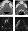
66-year-old female with poorly differentiated squamous cell carcinoma of the tongue. (a) Axial T1 weighted gadolinium-enhanced MRI with fat suppression through the level of the mandible and (b) coronal T2 weighted MRI with fat suppression show an enhancing mass (black arrow) with heterogeneous T2 signal (white arrow) in the left tongue invading into the superficial tongue muscles and genioglossus muscle, but with preservation of lingual septum. Invasion of extrinsic tongue musculature is considered T4 disease.
Figure 14
Figure 14
29-year-old female with a dermoid cyst in the root of tongue. (a) Axial T2 weighted MRI through the floor of mouth and (b) coronal post-contrast T1 with fat suppression MRI show an ovoid T2 hyperintense, non-enhancing mass (long arrow) that splays both genioglossus muscles (g). The mass also obstructs the right submandibular (Wharton's) duct (small arrow).
Figure 15
Figure 15
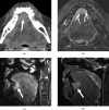
61-year-old female with poorly differentiated squamous cell carcinoma of the root of tongue. (a) Initial evaluation by contrast-enhanced CT at the level of the mandibular alveolus demonstrated an ill-defined hyperdense mass (arrow) in the anterior floor of mouth. (b) Further characterisation with axial T2 MRI at the same level demonstrates a well-defined T2 hyperintense mass in the anterior floor of mouth extending posteriorly to involve the genioglossus muscle. The mass obstructs the submandibular ducts bilaterally (small arrows). Sagittal (c) gadolinium-enhanced T1 and (d) fat-suppressed T2 weighted MRIs demonstrate the full extent of the anterior floor of mouth lesion (arrows), which is seen to involve the anterior fibres of the genioglossus muscle and the ventral surface of the tongue. This necessitates more complex resection and reconstructive surgery.
Figure 16
Figure 16
51-year-old male with acute hypoglossal denervation secondary to right internal carotid artery dissection. Initial presentation was of 1 week of dysarthria and hoarse voice. Subsequent clinical assessment of a right posterior tongue mass led to nasendoscopy and biopsy. Further evaluation with T1 weighted MRI through the floor of the mouth demonstrates (a) increased volume within the right posterior tongue with mass effect (arrow), (b) heterogeneous enhancement post contrast (s, submandibular gland) (arrows) and (c) subtle increased signal on T2 weighted images inkeeping with acute hypoglossal denervation. (d) Axial T1 weighted MRI through the level of the nasopharynx demonstrates the causative right internal carotid dissection (short arrow). Normal left internal carotid artery (thin arrow).
Figure 17
Figure 17
72-year-old male with hypoglossal denervation secondary to haemangiopericytoma. Axial contrast-enhanced CT images through the level of the (a) mandibular alveolus and (b) maxillary alveolus show fatty atrophy of the right tongue (arrow) involving the intrinsic and extrinsic muscles owing to a skull base haemangiopericytoma (open arrow) involving the right hypoglossal canal. (c) Axial and (d) coronal T1 weighted MRI shows well-demarcated T1 hyperintensity and volume loss in the intrinsic and extrinsic muscles of the right tongue (arrows). The normal left tongue has a “mass-like” appearance compared with the atrophic right side.
Figure 18
Figure 18
55-year-old female with a tumour involving the left lateral tongue. (a) The lesion is well depicted with intra-oral ultrasound (arrow). Clinical assessment suggested ipsilateral lymphadenopathy. (b) Coronal and (c) axial T2 weighted MRI with fat suppression through the level of the floor of mouth demonstrated the palpable abnormality to represent herniation of the sublingual gland through a defect in the mylohyoid muscle (short arrows). (d) This lesion is difficult to identify on CT (arrow).
Figure 19
Figure 19
56-year-old male with left buccal mucosa squamous cell carcinoma (SCC). (a) Post-contrast CT examination through the level of the mandibular neck and (b) coronal reformation. Note that the buccal mucosal SCC is easily discerned by puffed cheek CT (arrows). This technique requires patients to puff out their cheeks during CT scanning.
Figure 20
Figure 20
Differences in image quality obtained from angled gantry. (a) Routine axial CT of the oral cavity and (b) scout view with corresponding gantry angle demonstrates dental amalgam artefact obscuring anatomical detail. (c) and (d) angled gantry along plane of the mandible improves visualisation of the oral cavity by reducing dental amalgam artefact. Note concurrent use of puff cheek technique.
Figure 21
Figure 21
57-year-old man with right tongue squamous cell carcinoma. (a) Coronal T2 weighted MRI with fat saturation, the tongue tumour (arrow) is poorly delineated with the depth of invasion difficult to ascertain. (b) Longitudinal ultrasound image of the right lateral tongue obtained with an intra-oral probe. There is excellent delineation of the hypoechoic tumour relative to the hyperechoic intrinsic tongue musculature.

References

    1. MacDonald AJ, Harnsberger HR. Oral cavity anatomy and imaging issues. Harnsberger HR, Wiggins RH, Hudgins PA, Michel MA, Swartz J, Davidson HC, et al.,eds. Diagnostic imaging: head and neck. Salt Lake City, UT: Amirsys; 2004;III-:42–5
    1. Smoker WRK. The oral cavity. Som PM, Curtin HD, eds. Head and neck imaging, 4th edn. St Louis, MO. Mosby: 2003:1377–1464
    1. Harnsberger HR. Saint Louis, MO: Mosby: 1995. Handbook of head and neck imaging. 2nd edn.
    1. McMinn RMH, ed. Last's anatomy: regional and applied. New York, NY, USA, Churchill Livingstone; 1994
    1. Stambuk HE, Karimi S, Lee N, Patel SG. Oral cavity and oropharynx tumours. Radiol Clin N Am 2007;45:1–20 - PubMed