. 2011;6(9):e23348.
doi: 10.1371/journal.pone.0023348. Epub 2011 Sep 14.

Human ovarian tumor cells escape γδ T cell recognition partly by down regulating surface expression of MICA and limiting cell cycle related molecules

Affiliations

Human ovarian tumor cells escape γδ T cell recognition partly by down regulating surface expression of MICA and limiting cell cycle related molecules

Jingwei Lu et al. PLoS One. 2011.

Abstract

Background: Mechanisms of human Vγ2Vδ2 T cell-mediated tumor immunity have yet to be fully elucidated.

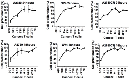
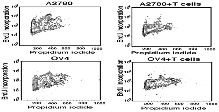
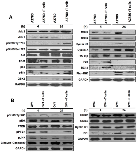
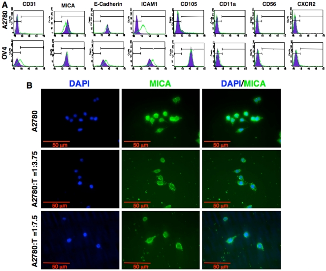
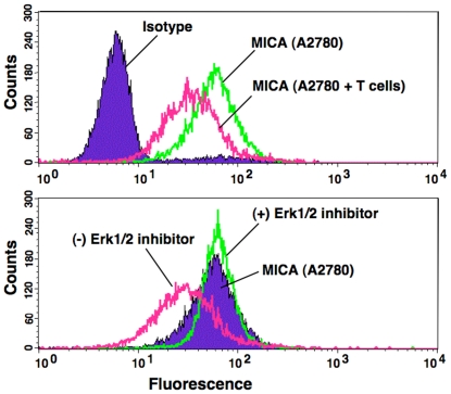
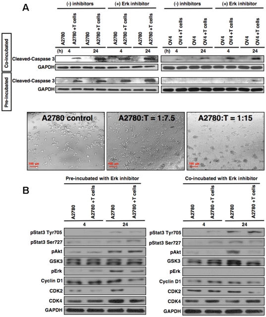
Methods and findings: At least some tumor cell recognition is mediated by NKG2D-MICA interactions. Herein, by using MTT assay and PI-BrdU co-staining and Western-blot, we show that these Vγ2Vδ2 T cells can limit the proliferation of ovarian tumor cells by down regulation of apoptosis and cell cycle related molecules in tumor cells. Cell-to-cell contact is critical. γδ T cell-resistant, but not susceptible ovarian tumor cells escape γδ T cell-mediated immune recognition by up-regulating pErk1/2, thereby decreasing surface MICA levels. Erk1/2 inhibitor pretreatment or incubation prevents this MICA decrease, while up-regulating key cell cycle related molecules such as CDK2, CDK4 and Cyclin D1, as well as apoptosis related molecules making resistant tumor cells now vulnerable to γδ T cell-mediated lysis.

Conclusion: These findings demonstrate novel effects of γδT cells on ovarian tumor cells.


Conflict of interest statement

Competing Interests: The authors have declared that no competing interests exist.

Figures

Figure 1
Figure 1. Tumor apoptosis mediated by γδ T cells.
A. Morphology of tumor cells after co-culture with γδ T cells. Various ovarian tumor cell lines stated (A2780, OV4 and A2780CR) were co-cultured with γδ T cells for 24 hours with a ratio of Cancer∶T = 1∶0, 1∶7.5 or 1∶15 and morphology of tumor cells after co-culture with γδ T cells were captured by phase contrast micrographic images. Detached tumor cells formed clumps along with γδ T cells. Five experiments were performed using expanded γδ T cells isolated from three different donors. Results were similar and representative data is shown here. B. Flowcytometric analyses for apoptosis marker, annexin V, were performed after co-culture of tumor cells (A2780 or OV4) with γδ T cells (1∶10 ratio) for 24 h. The expression of annexin V was much higher in OV4 cells compared to A2780 cells, indicating enhanced apoptosis in OV4 cells. C. Higher cell death in OV4 line was also correlated with the higher level of cleaved caspase 3 protein evaluated by Western blot analysis.
Figure 2
Figure 2. Proliferation of tumor cells in presence of γδ T cells.
Ovarian tumor cell lines (A2780, OV4 and A2780CR) were co-cultured with various ratios of γδ T cells for 24 and 48 hours. MTT assay was performed to evaluate cell proliferation after gentle removal of γδ T cells after 24 and 48 hours of co-culture.
Figure 3
Figure 3. BrdU incorporation and propidium iodide (PI) co-staining in tumor cells.
Ovarian tumor cells, A2780 and OV4 were co-cultured for 24 h in the presence or absence of γδ T cells at the ratio of 1∶15. After co-culture, cells were pulsed with BrdU for 5 hours and PI staining was performed prior to flowcytometric analyses.
Figure 4
Figure 4. Expression of signaling molecules on tumor cells.
Ovarian tumor cell lines were co-culturing with γδ T cells for 4 hours and 24 hours at 1∶5 ratio. After gentle removal of γδ T cells proteins were harvested from tumor cells and Western blot was performed for various molecules stated in A2780 (A) and OV4 (B) cells.
Figure 5
Figure 5. Expression of cell surface molecules on ovarian tumor cells after co-culture with γδ T cells.
A. Flowcytometric analysis of various surface markers on tumor cells after co-culture with γδ T cells (1∶7.5 ratio) for 24 h. B. Expression of MICA on A2780 cells was evaluated by using immunocytochemical staining after co-culture with various concentration of γδ T cells.
Figure 6
Figure 6. Surface expression of MICA on A2780 cells after co-culture with γδ T cells or Erk1/2 inhibitor.
A2780 cells were co-cultured with γδ T cells (1∶7.5 ratio) for 36 h and flowcytometric analysis was performed to determine the level of surface expression of MICA on 2780 cells (upper panel). A2780 cells were co-cultured with γδ T cells for 24 h and gently removed the γδ T cells. A2780 cells were then cultured for another 12 h in presence or absence of Erk1/2 inhibitor. Flowcytometric analysis was performed for MICA expression (lower panel).
Figure 7
Figure 7. Effect of Erk1/2 inhibitor on tumor cell morphology and levels of signaling molecules after γδ T cells co-culture.
A. A2780 or OV4 cells were pre-incubated with Erk inhibitor (−/+) for one hour or co-incubated with Erk inhibitors (−/+) until the termination of experiment. Cells were then co-cultured with γδ T cells (1∶5 ratio), and at various time points A2780 cells or OV4 cells were harvested after gentle removal of γδ T cells. Protein related to apoptosis (upper panel) was analyzed using Western blot. Cell morphology was determined under a phase contrast microscope after 1 hour of Erk-inhibitor pulse followed by 24 h of co-culture with γδ T cells at various ratios (lower panel). B. A2780 cells were pre-incubated with Erk inhibitor for one hour and then co-cultured with γδ T cells (1∶5 ratio) or co-incubated with Erk inhibitor during co-culture with γδ T cells and at various time points A2780 cells were harvested after gentle removal of γδ T cells. Proteins related to cell cycles and proliferations were analyzed using Western blot.

References

    1. Porcelli S, Brenner MB, Band H. Biology of the human gamma delta T-cell receptor. Immunol Rev. 1991;120:137–183. - PubMed
    1. Morita CT, Parker CM, Brenner MB, Band H. TCR usage and functional capabilities of human gamma delta T cells at birth. J Immunol. 1994;153:3979–3988. - PubMed
    1. Hara T, Mizuno Y, Takaki K, Takada H, Akeda H, et al. Predominant activation and expansion of V gamma 9-bearing gamma delta T cells in vivo as well as in vitro in Salmonella infection. J Clin Invest. 1992;90:204–210. - PMC - PubMed
    1. Bukowski JF, Morita CT, Brenner MB. Human gamma delta T cells recognize alkylamines derived from microbes, edible plants, and tea: implications for innate immunity. Immunity. 1999;11:57–65. - PubMed
    1. Morita CT, Jin CG, Sarikonda G, Wang H. Nonpeptide antigens, presentation mechanisms, and immunological memory of human V gamma 2 V delta 2 T cells: discriminating friend from foe through the recognition of prenyl pyrophosphate antigens. Immunological Reviews. 2007;215:59–76. - PubMed

Publication types

MeSH terms

Substances