. 2011;6(9):e24080.
doi: 10.1371/journal.pone.0024080. Epub 2011 Sep 15.

The phenotypic radiation resistance of CD44+/CD24(-or low) breast cancer cells is mediated through the enhanced activation of ATM signaling

Affiliations

The phenotypic radiation resistance of CD44+/CD24(-or low) breast cancer cells is mediated through the enhanced activation of ATM signaling

Hong Yin et al. PLoS One. 2011.

Abstract

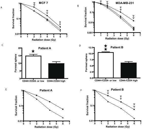
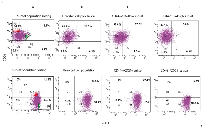
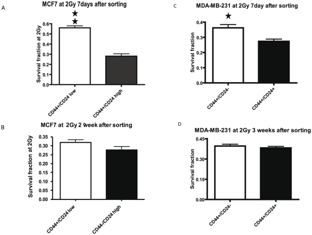
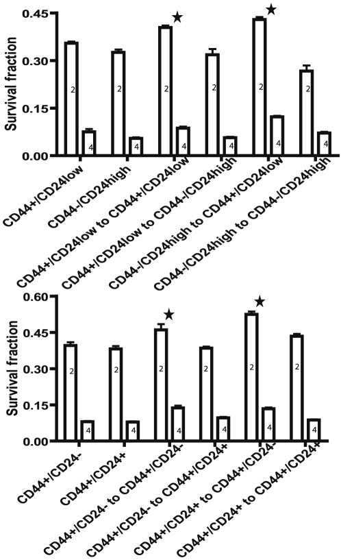
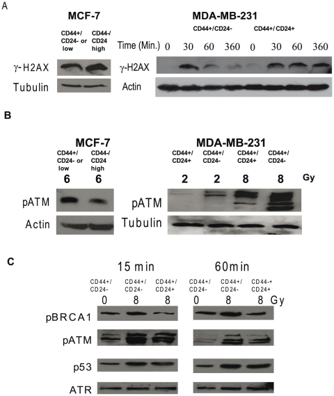
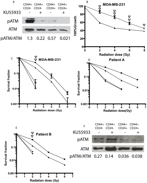
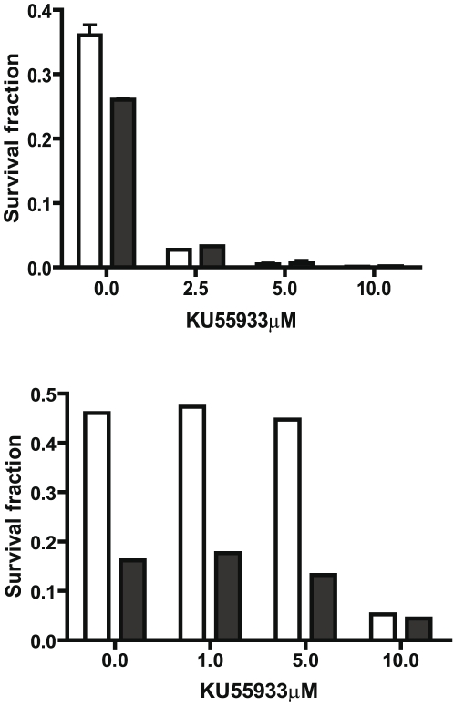
Cancer initiating cells (CIC) are stem-like cells. CIC may contribute not only to the initiation of cancer but also to cancer recurrence because of the resistance of CIC both to chemotherapy and radiation therapy. From the MCF-7 and MDA-MB231 breast cancer cell lines and primary culture of patient breast cancer cells, we isolated by flow cytometry a CIC subset of cells with the CD44(+)/CD24(-or low) phenotype. The CD44(+)/CD24(-or low) subset showed increased sphere formation and resistance to radiation compared to the non- CD44(+)/CD24(-or low) subset. The increased radiation resistance was not dependent on the result of altered non-homologous end joining (NHEJ) DNA repair activity as both NHEJ activity and expression of the various proteins involved in NHEJ were not significantly different between the CD44(+)/CD24(-or low) and non- CD44(+)/CD24(-or low) subsets. However, activation of ATM signaling was significantly increased in CD44(+)/CD24(-or low) cells compared to non- CD44(+)/CD24(-or low) cells in both from breast cancer cell lines and primary human breast cancer cells. Application of an ATM inhibitor effectively decreased the radiation resistance of CD44(+)/CD24(-or low) subset, suggesting that targeting ATM signaling may provide a new tool to eradicate stem-like CIC and abolish the radiation resistance of breast cancer.


Conflict of interest statement

Competing Interests: The authors have declared that no competing interests exist.

Figures

Figure 1
Figure 1. Post-radiation clonogenic survival and mammosphere formation analyses of the CD44+/CD24−or low subset vs. CD44/CD24high (MCF-7, Patient A, and Patient B) and CD44+/CD24+ (MDA-MB-231) subsets.
A and B. As detailed in the Methods radiation dose-response curves were performed either of unsorted MCF-7 cells (▾) or the MCF-7 cell subsets CD44+/CD24−or low (▪) and CD44/CD24high (▴) (Panel A) or unsorted MDA-MB-231 cells (▾), or the MDA-MB-231 cell subsets CD44+/CD24 (▪) or CD44+/CD24+ (▴) (Panel B). Double open triangles indicate a significant difference at the indicated dose of radiation with a P<0.01 between the CD44+/CD24−or low subset and the CD44/CD24high subset or unsorted cells for MCF-7 cells and between the CD44+/CD24 subset and the CD44+/CD24+ subset or unsorted cells for MDA-MB-231 cells. C. and D. Cells were cultured to form mammospheres from the CD44+/CD24−or low or the CD44+/CD24high subsets of Patient A (Panel C) and Patient B (Panel D) as detailed in the Methods. In brief, the cells sorted from primary cultures were grown on low cell binding 96 well plates at a density of 200 cells in 0.2 ml of medium per well. The spheres formed were counted at day 12. The * indicates a significant difference with P<0.05 and ** indicates a significant difference with P<0.01 between the CD44+/CD24−or low subset and CD44/CD24highsubset. E and F. A radiation dose response curve was performed on the CD44+/CD24−or low subset (▪) and the CD44+/CD24+ subset (▴) from Patient A (Panel E) and Patient B (Panel F) as detailed in the Methods. Double open triangles indicate a significant difference at the indicated radiation dose with P<0.01 between the CD44+/CD24−or low subset and the CD44/CD24high subset in patient B cells.
Figure 2
Figure 2. CD44/CD24 expression after culture of sorted cells.
MCF-7 (top panel) and MDA-MB-231 (bottom panel) cells were sorted by the surface markers CD44 and CD24 as shown (column A) into CD44+/CD24−or low (3.0%) and CD44/CD24high (3.2%) subsets for MCF-7 or CD44+/CD24 (4.5%) and CD44+/CD24+ (4.5%) for MDA-MB-231. The parent cell lines (column B) and the subsets (columns C and D) were cultured for 20 days and re-examined for CD44 and CD24 expression. The fluorescent intensity of CD 24 expression is on the Y axis and of CD44 expression on the X axis. Shown is the percentage of cells in each quadrant.
Figure 3
Figure 3. The affect of culture on the radiation resistance of stem-like subsets.
MCF-7 cells (Panels A and B) were sorted into CD44+/CD24low (open bars) and CD44/CD24high (solid bars) subsets. MDA-MB-231 cells (Panels C and D) were sorted into CD44+/CD24 (open bars) and CD44+/CD24+ (solid bars) subsets. A clonogenic survival analysis was conducted with calculation of the survival fraction at 2 Gy of radiation after 7 days (A and C), two weeks (B), or three weeks (D) of culture. Each column represents the mean of three independent experiments ± SD. The single and double stars represent a significant difference between two subsets in MCF7 and MDA-MB-231 cells with P<0.05 and P<0.01, respectively.
Figure 4
Figure 4. Analysis of radiation resistance of stem-like subsets upon initial isolation and on the same subset obtained upon resorting after culture.
MCF7 cells (upper panel) were sorted into CD44+/CD24low and CD44/CD24high subsets and MDA-MB-231 cells (lower panel) were sorted into CD44+/CD 24 and CD44+/CD 24+ subsets. All four subsets were cultured for 20 days, resorted into the same subsets and clonogenic survival analyses were performed after radiation with either 2 Gy (2) or 4 Gy (4). Shown are the means ± SD of 3 experiments. Statistical analyses were performed with two-way ANOVA analysis. The stars indicate a significant difference with P<0.01 in the survival fraction of the resorted CIC group compared to the resorted non CIC groups. In upper panel CD44+/CD24low and CD44/CD24high mean 20 day cultured MCF7 subsets. CD44+/CD24low to CD44+/CD24low and CD44+/CD24low to CD44/CD24high indicate that cells were re-sorted into CD44+/CD24low and CD44/CD24high subsets from 20 day cultured CD44+/CD24low subset. CD44/CD24high to CD44+/CD24lowand CD44/CD24high to CD44/CD24high indicate that cells were re-sorted into CD44+/CD24low and CD44/CD24high subsets from 20 day cultured CD44/CD24high subset. In lower panel CD44+/CD24 and CD44+/CD24+ mean 20 day cultured MDA-MB-231 subsets. CD44+/CD24 to CD44+/CD24 and CD44+/CD24 to CD44+/CD24+ indicate that cells were re-sorted into CD44+/CD24 and CD44+/CD24+ subsets from 20 day cultured CD44+/CD24 subset. CD44+/CD24+ to CD44+/CD24and CD44+/CD24+ to CD44+/CD24+ indicate that cells were re-sorted into CD44+/CD24 and CD44+/CD24+ subsets from 20 day cultured CD44+/CD24+ subset.
Figure 5
Figure 5. The differential response to radiation correlates with differential phosphorylation of ATM and its downstream targets between CD44+/CD24 and CD44+/CD24+ subsets in MDA-MB231 cells and between CD44+/CD24low and CD44/CD24high subsets in MCF-7 cells.
The indicated sorted subsets of MCF-7 and MDA-MB231 cells were cultured for 5 days as described in the Methods and radiated with the indicated doses. Whole cell extracts (MCF-7) or nuclear extracts (MDA-MB-231) were analyzed by western blot analysis for the presence of phosphorylated γH2AX (A), phosphorylated ATM (B), and BRCA1 and p53 (C) the downstream targets of phosphorylated ATM. between CD44+/CD24 and CD44+/CD24+ subsets of MDA-MB231 cells. In C western blot analyses were conducted for time-related changes in phosphorylated ATM, phosphorylated BRCA1, and phosphorylated p53. The ATR expression was utilized as a loading control.
Figure 6
Figure 6. The ATM inhibitor KU55933 decreases the radiation resistance of stem-like MDA-MB-231, Patient A, and Patient B cells.
Cells sorted and cultured as in Figure 1 were treated with ATM inhibitor KU55933. A. Western blot analysis of phosphorylated ATM 30 minutes after radiation at 10Gy in MDA-MB-231 cells treated with 20 µM of the ATM inhibitor KU55933 for two hours before radiation. B. The proliferation analysis of ATM inhibitor treated cells. MDA-MB-231 cells were treated with the 20 µM of KU55933 from two hours before radiation to 24 hour after radiation and cell numbers enumerated as described in the Methods after 7 days in culture. The results are expressed as the percent of cells compared to the non-radiated control and represent the mean ± SD of three independent experiments. The solid squares represent the CD44+/CD24 subset, the triangles, the CD44+/CD24+ subset. Single and double open triangles indicate a significant difference with P<0.05 and 0.01 between KU55933 treated and untreated CD44+/CD24 subset of MDA-MB-231 cells. C, D, and E. The clonogenic survival analysis of the CD44+/CD24 subset vs. CD44+/CD24+ subset in MDA-MB-231 cells (C) and CD44+/CD24−or low subset vs CD44/CD24high subset in Patient A (D) and Patient B cells (E). Sorted cells were treated with 10 µM of KU55933 one hour before the radiation and then irradiated as described in Materials and Methods. 48 hour after radiation, medium was replaced with fresh medium without KU55933. The colonies were counted at day 12. The solid square represents the CD44+/CD24 subset in C and CD44+/CD24−or low subset in D and E. The ▴ triangle represents the CD44+/CD24+ subset in C and the CD44/CD24high subset in D and E. The ▾ triangle represents the KU55933 treated CD44+/CD24 subset in C and CD44+/CD24−or low subset in D and E. The ♦ represents KU55933 treated CD44+/CD24+ subset in C and CD44/CD24high subset in D and E. Double open triangles indicate a significant difference at 2Gy with P<0.01 between KU55933 treated and untreated CD44+/CD24 subsets in C and between the KU55933 treated and untreated CD44+/CD24−or low subset in E. F. Western blot analysis of phosphorylated ATM 60 minutes after radiation at 6 Gy in Patient B cells treated with 10 µM of the ATM inhibitor KU55933 for one hour prior to radiation.
Figure 7
Figure 7. Dose-effect relationship of KU55933 and clonogenic survival fraction in MDA-MB-231 and Patient B cells.
Sorted MDA-MB-231 (A) and patient B (B) cells were treated with various concentrations of KU55933 as indicated for one hour before the radiation and then irradiated at 2Gy. The colonies were enumerated as in Figure 6 and the survival fractions were calculated The open bar represents the CD44+/CD24 subset in A and the CD44+/CD24−or low subset in B. The solid bar represents the CD44+/CD24+ subset in A and CD44/CD24high subset in B.

References

    1. Ponti D, Zaffaroni N, Capelli C, Daidone MG. Breast cancer stem cells: an overview. Eur J Cancer. 2006;42:1219–1224. - PubMed
    1. Woodward WA, Chen MS, Behbod F, Rosen JM. On mammary stem cells. J Cell Sci. 2005;118:3585–3594. - PubMed
    1. Al-Hajj M, Wicha MS, Benito-Hernandez A, Morrison SJ, Clarke MF. Prospective identification of tumorigenic breast cancer cells. Proc Natl Acad Sci U S A. 2003;100:3983–3988. - PMC - PubMed
    1. Shipitsin M, Campbell LL, Argani P, Weremowicz S, Bloushtain-Qimron N, et al. Molecular definition of breast tumor heterogeneity. Cancer Cell. 2007;11:259–273. - PubMed
    1. Giatromanolaki A, Sivridis E, Fiska A, Koukourakis MI. The CD44+/CD24− phenotype relates to ‘triple-negative’ state and unfavorable prognosis in breast cancer patients. Med Oncol. 2011;28:745–752. - PubMed

Publication types

MeSH terms