

Humanized mouse model of ovarian cancer recapitulates patient solid tumor progression, ascites formation, and metastasis
- PMID: 21935406
- PMCID: PMC3174163
- DOI: 10.1371/journal.pone.0024420
Humanized mouse model of ovarian cancer recapitulates patient solid tumor progression, ascites formation, and metastasis
Abstract
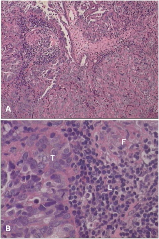
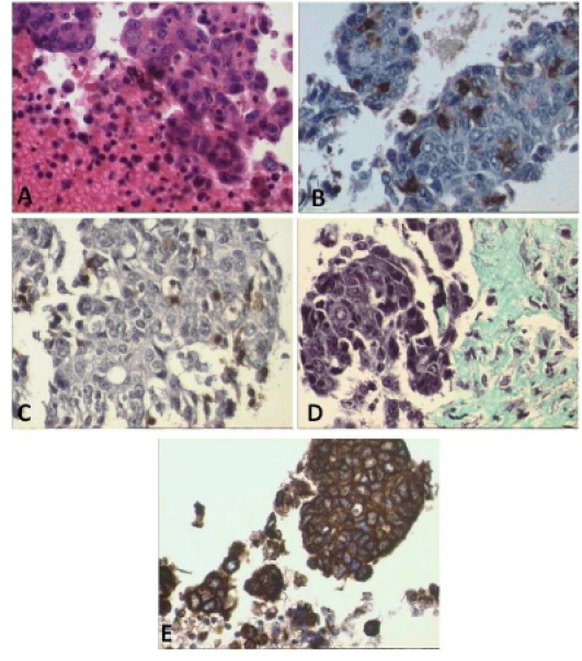
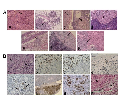
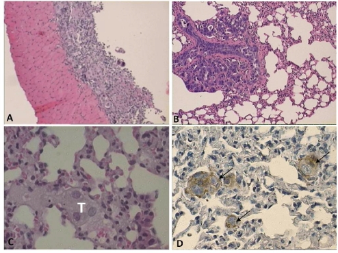
Ovarian cancer is the most common cause of death from gynecological cancer. Understanding the biology of this disease, particularly how tumor-associated lymphocytes and fibroblasts contribute to the progression and metastasis of the tumor, has been impeded by the lack of a suitable tumor xenograft model. We report a simple and reproducible system in which the tumor and tumor stroma are successfully engrafted into NOD-scid IL2Rγ(null) (NSG) mice. This is achieved by injecting tumor cell aggregates derived from fresh ovarian tumor biopsy tissues (including tumor cells, and tumor-associated lymphocytes and fibroblasts) i.p. into NSG mice. Tumor progression in these mice closely parallels many of the events that are observed in ovarian cancer patients. Tumors establish in the omentum, ovaries, liver, spleen, uterus, and pancreas. Tumor growth is initially very slow and progressive within the peritoneal cavity with an ultimate development of tumor ascites, spontaneous metastasis to the lung, increasing serum and ascites levels of CA125, and the retention of tumor-associated human fibroblasts and lymphocytes that remain functional and responsive to cytokines for prolonged periods. With this model one will be able to determine how fibroblasts and lymphocytes within the tumor microenvironment may contribute to tumor growth and metastasis, and will make it possible to evaluate the efficacy of therapies that are designed to target these cells in the tumor stroma.
Conflict of interest statement
Figures
References
-
- Reddy S, Piccione D, Takita H, Bankert RB. Human lung tumor growth established in the lung and subcutaneous tissue of mice with severe combined immunodeficiency. Cancer Res. 1987;47:2456–2460. - PubMed
-
- Shultz LD, Ishikawa F, Greiner DL. Humanized mice in translational biomedical research. Nature Rev Immunol. 2007;7:118–130. - PubMed
-
- Bankert RB, Egilmez NK, Hess SD. Human-SCID mouse chimeric models for the evaluation of anti-cancer therapies. TRENDS in Immunol. 2001;22:386–393. - PubMed
-
- Erez N, Truitt M, Olson P, Hanahan D. Cancer-associated fibroblasts are activated in incipient neoplasia to orchestrate tumor-promoting inflammation in an NF-kappaB-dependent manner. Cancer Cell. 2010;17:135–147. - PubMed
-
- Simpson-Abelson MR, Sonnenberg GF, Takita H, Yokota SJ, Conway TJ, Jr, et al. Long-term engraftment and expansion of tumor-derived memory T cells following the implantation of non-disrupted pieces of human lung tumor into NOD-scid IL2Rγnull mice. J Immunol. 2008;180:7009–7018. - PubMed
Publication types
MeSH terms
Substances
Grants and funding
LinkOut - more resources
Full Text Sources
Other Literature Sources
Medical
Molecular Biology Databases
Research Materials
Miscellaneous
