. 2011;6(9):e24656.
doi: 10.1371/journal.pone.0024656. Epub 2011 Sep 14.

Acute drug treatment in the early C. elegans embryo

Affiliations

Acute drug treatment in the early C. elegans embryo

Ana Carvalho et al. PLoS One. 2011.

Abstract

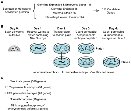
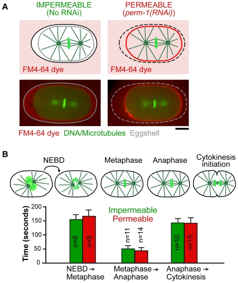
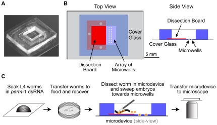
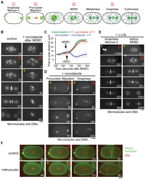
Genetic and genome-wide RNAi approaches available in C. elegans, combined with tools for visualizing subcellular events with high-resolution, have led to increasing adoption of the early C. elegans embryo as a model for mechanistic and functional genomic analysis of cellular processes. However, a limitation of this system has been the impermeability of the embryo eggshell, which has prevented the routine use of small molecule inhibitors. Here, we present a method to permeabilize and immobilize embryos for acute inhibitor treatment in conjunction with live imaging. To identify a means to permeabilize the eggshell, we used a dye uptake assay to screen a set of 310 candidate genes defined by a combination of bioinformatic criteria. This screen identified 20 genes whose inhibition resulted in >75% eggshell permeability, and 3 that permeabilized embryos with minimal deleterious effects on embryo production and early embryonic development. To mount permeabilized embryos for acute drug addition in conjunction with live imaging, we combined optimized inhibition of one of these genes with the use of a microfabricated chamber that we designed. We demonstrate that these two developments enable the temporally controlled introduction of inhibitors for mechanistic studies. This method should also open new avenues of investigation by allowing profiling and specificity-testing of inhibitors through comparison with genome-wide phenotypic datasets.


Conflict of interest statement

Competing Interests: The authors have declared that no competing interests exist.

Figures

Figure 1
Figure 1. A screen for genes required for eggshell impermeability.
A. Since the eggshell is a secreted structure, all candidate genes encoded proteins with a signal peptide or a membrane-targeting domain. Our candidate gene set included all of the germline enriched ([13], [14]) and maternal sterile ([5]) genes that have a signal peptide. Since the set of genes expressed in dissected germlines that have a signal peptide is large (1137 genes, Serial Analysis of Gene Expression database, Genome BC C. elegans Gene Expression Consortium, http://elegans.bcgsc.bc.ca) we included only those previously shown to result in embryonic lethality when inhibited by RNAi (Wormbase release WS199). Genes with proteins domains (chitin binding, glycosylation, protease, peroxidase, lipid binding, or LDLR domains) found in proteins implicated in eggshell formation or analogous processes in other species were included regardless of whether there was prior evidence for a germline role. Due to redundancy, the total number of candidate genes was 310. B. Outline of screen to identify genes specifically required for eggshell impermeability. C. Of the 310 candidate genes tested, 3 genes were identified whose inhibition permeabilized the eggshell with high penetrance (>75% permeable embryos), while maintaining a normal broodsize and leading to minimal defects in gonad structure/embryonic development. One of these genes, perm-1, was chosen for further optimization.
Figure 2
Figure 2. perm-1 inhibition permeabilizes the eggshell without altering cell division timing.
A. Schematics and confocal images of impermeable (no RNAi) and permeable (perm-1(RNAi)) embryos co-expressing GFP-histone H2B and GFP-alpha-tubulin. Embryos at metaphase of the first mitotic division were submerged in media containing the lipophilic dye FM4-64, which passes through the eggshell to label the plasma membrane of permeable (right), but not impermeable (left) embryos. Scale bar, 10 µm. B. Time intervals between NEBD and metaphase, metaphase and anaphase, and anaphase and cytokinesis onset were measured for permeable embryos (perm-1(RNAi)); red bars) and impermeable wild-type controls (green bars).
Figure 3
Figure 3. A microdevice that immobilizes permeable embryos for simultaneous imaging and small molecule addition.
A. Photograph of the microdevice. B. Schematic drawings of the top and side views of the microdevice. C. Schematic depicting the procedure for preparing permeabilized embryos for drug treatment and imaging. C. elegans hermaphrodites at the L4 larval stage are soaked in a drop of perm-1 dsRNA for 4 hours at 20°C. The worms are transferred to a plate with bacteria and allowed to recover for 16 hours at 16°C. 1–3 worms are placed on the dissection board of the microdevice after filling it with ∼100 µl of media. Worms are dissected with a scalpel and the released early embryos are swept into the microwells with an eyelash tool. The microdevice is transferred to the microscope and the embryos are imaged. When the desired stage is reached, the medium in the microdevice is replaced with fresh medium containing the drug of interest.
Figure 4
Figure 4. Acute drug treatments with nocodazole, latrunculin A, and c-lactocystin-ß-lactone.
A. Schematics illustrating the stages between anaphase meiosis II and cytokinesis of the first mitotic division following fertilization. Red numbers mark the timepoints highlighted in the panels in B, D, E and F. B. Confocal images of permeable embryos expressing GFP-histone H2B and GFP-alpha-tubulin. A control embryo (left) and an embryo treated with 10 µg/ml nocodazole after NEBD (right) are shown. Addition of nocodazole results in rapid microtubule depolymerization. Numbers in white indicate time in seconds after NEBD. Scale bar, 5 µm. C. Mean pole-to-pole distance plotted versus time for the indicated conditions. Embryos treated with nocodazole after NEBD exhibit a rapid reduction in pole-to-pole distance due to microtubule depolymerization. Error bars are the 95% C.I. of the mean. D. Images of permeable embryos expressing GFP-histone and GFP-alpha-tubulin. Nocodazole was added during pronuclear migration (left) or in early anaphase (right). In both cases, rapid microtubule depolymerization was observed. Scale bar, 10 µm. Numbers in white indicate time in seconds after the timepoint when drug was added. E. Images of permeable embryos expressing GFP-histone H2B and GFP-alpha-tubulin. The proteasome inhibitor, c-lactocystin-ß-lactone (20 µM) was added either early (anaphase of meiosis II; left) or just before NEBD (right). If added early, the embryo arrested at the first metaphase. If added before NEBD, the metaphase-anaphase transition was delayed, but the embryo did not arrest until metaphase of the second division. Scale bar, 5 µm. Numbers in white indicate time in seconds after NEBD of the first mitotic division. F. Images of embryos expressing a GFP-labeled plasma membrane probe and RFPmCherry-histone H2B. Latrunculin A (10 µM) added after anaphase onset prevented cleavage furrow ingression. Scale bar, 10 µm. Numbers in white indicate time in seconds after anaphase onset. Yellow asterisks in the upper left corner of the panels in B, D, E and F mark the time point when drug was added.

References

    1. Hyman AA, White JG. Determination of cell division axes in the early embryogenesis of Caenorhabditis elegans. J Cell Biol. 1987;105:2123–2135. - PMC - PubMed
    1. Hill DP, Strome S. Brief cytochalasin-induced disruption of microfilaments during a critical interval in 1-cell C. elegans embryos alters the partitioning of developmental instructions to the 2-cell embryo. Development. 1990;108:159–172. - PubMed
    1. Edgar LG. Blastomere culture and analysis. Methods Cell Biol. 1995;48:303–321. - PubMed
    1. Rappleye CA, Tagawa A, Lyczak R, Bowerman B, Aroian RV. The anaphase-promoting complex and separin are required for embryonic anterior-posterior axis formation. Dev Cell. 2002;2:195–206. - PubMed
    1. Green RA, Kao HL, Audhya A, Arur S, Mayers JR, et al. A High-Resolution C. elegans Essential Gene Network Based on Phenotypic Profiling of a Complex Tissue. Cell. 2011;145:470–482. - PMC - PubMed

Publication types

MeSH terms

LinkOut - more resources