. 2011;6(9):e24659.
doi: 10.1371/journal.pone.0024659. Epub 2011 Sep 15.

Pax6 represses androgen receptor-mediated transactivation by inhibiting recruitment of the coactivator SPBP

Affiliations

Pax6 represses androgen receptor-mediated transactivation by inhibiting recruitment of the coactivator SPBP

Julianne Elvenes et al. PLoS One. 2011.

Abstract

The androgen receptor (AR) has a central role in development and maintenance of the male reproductive system and in the etiology of prostate cancer. The transcription factor Pax6 has recently been reported to act as a repressor of AR and to be hypermethylated in prostate cancer cells. SPBP is a transcriptional regulator that previously has been shown to enhance the activity of Pax6. In this study we have identified SPBP to act as a transcriptional coactivator of AR. We also show that Pax6 inhibits SPBP-mediated enhancement of AR activity on the AR target gene probasin promoter, a repression that was partly reversed by increased expression of SPBP. Enhanced expression of Pax6 reduced the amount of SPBP associated with the probasin promoter when assayed by ChIP in HeLa cells. We mapped the interaction between both AR and SPBP, and AR and Pax6 to the DNA-binding domains of the involved proteins. Further binding studies revealed that Pax6 and SPBP compete for binding to AR. These results suggest that Pax6 represses AR activity by displacing and/or inhibiting recruitment of coactivators to AR target promoters. Understanding the mechanism for inhibition of AR coactivators can give rise to molecular targeted drugs for treatment of prostate cancer.


Conflict of interest statement

Competing Interests: The authors have declared that no competing interests exist.

Figures

Figure 1
Figure 1. Pax6 represses SPBP-mediated enhancement of AR activity.
(A) Pax6 represses AR-mediated expression from the 285PB-Luc reporter. LNCaP cells were cultured in medium with charcoal treated serum and cotransfected with 5–200 ng pDestHA-Pax6, 75 ng pDestHA-AR, 75 ng p285PB-Luc, and 5 ng pCMV-βgal using Metafectene Pro (Biontex). pcDNA3HA was used to equalize the amount of DNA in each well. 10−7 M synthetic androgen (R1881) was added as indicated. The mean luciferase/β galactosidase value of AR-mediated expression from 285PB-Luc after stimulation with R1881 was set to 1. The data is based on the mean values of three independent experiments performed in triplicate. (B) SPBP enhances AR-mediated expression, a coactivation that is lost by coexpression of Pax6. HEK293 cells were cultured in medium with charcoal treated serum and cotransfected with 75 ng pDestHA-AR, 50 or 150 ng pDestHA-SPBP, 0–25 ng pDestHA-Pax6, 75 ng p285PB-Luc, and 5 ng pCMV-βgal. The experiment was performed and data obtained as described in A. (C) Chromatin Immunoprecipitation assays show that both SPBP and AR associate with the probasin promoter, and that overexpression of Pax6 decreases the amount of SPBP associated with the promoter. Extracts from HeLa cells cotransfected with 285PB-Luc, pDestHA-SPBP, pSG5-AR, and/or increasing amounts of pDestHA-Pax6 were immunoprecipitated with preimmune serum, polyclonal anti-SPBP antibody or polyclonal anti-AR antibody. PCR analyses on the immunoprecipitated chromatin were carried out using primers flanking the probasin promoter (upper panel). Primers aligning to position -3351 and -3069 of the cathepsin D promoter were used as control (lower panel).The 1 kb DNA ladder is shown to the left. (D) SiRNA knock down of endogenous SPBP in R1881 stimulated LNCaP cells reduces the expression of the AR target gene PSA. LNCaP cells were transfected with SPBP siRNA or scrambled siRNA, stimulated with R1881 for 48 hours before harvesting. RT-PCR reactions were run on PSA and β-actin mRNA. The average amount of PSA mRNA correlated to β-actin mRNA based on four independent experiments are shown with standard deviations. The right panel shows the knock-down of SPBP expression in LNCaP cells transfected with SPBP siRNA compared to scramled siRNA transfected and untransfected LNCaP cells.
Figure 2
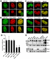
Figure 2. Colocalization and coexpression of AR, Pax6 and SPBP.
(A) and (B) Colocalization between GFP-AR and Cherry-Pax6 or Cherry-SPBP, respectively. The GFP-AR 3108 expressing cell line was transiently transfected with 100 ng pDestCherry-Pax6 or 150 ng pDestCherry-SPBP using TransIT-LT1 (Mirus Bio). The cells were stimulated with synthetic androgen R1881 and live cell images obtained using a Zeiss LSM510 confocal laser scanning microscope. Scale bars: 10 µm. (C) Colocalization between GFP- and Cherry-tagged SPBP and Pax6. HeLa cells were transiently cotransfected with either 175 ng pDestEGFP-SPBP and 25 ng pDestCherry-Pax6, or 25 ng pDestEGFP-Pax6 and 175 ng pDestCherry-SPBP using TransIT-LT1 (Mirus Bio). Live cell images were obtained as in A. Scale bars: 10 µm. (D) Pearson's correlation coefficients support the colocalization of GFP-AR with mCherry-SPBP and mCherry-Pax6 shown in A and B. The correlation is based on the average of 5–10 independent cells, with standard deviations shown. The nuclear correlation between mCherry-Pax6 and nuclear GFP, and GFP-Pax6 and nuclear mCherry, were used as negative controls. (E) Coexpression of Pax6, SPBP and AR in mammalian cell lines. Approximately 50 µg proteins from each of the whole cell extracts were separated on 6 and 12% SDS-polyacrylamide gels, followed by immunoblotting using anti-Pax6 (Chemicon), anti-AR (Santa Cruz), anti-SPBP and anti-actin (Sigma) antibodies. Actin was used as a loading control. The cell lines used were HeLa (human epithelial), HEK293 (human embryonic kidney), MEF (mouse embryonic fibroblast), Kelly and SKNBe(2) (human neuroblastoma), and Du145, PC3 and LNCaP (human prostate cancer).
Figure 3
Figure 3. Pax6 and AR interact directly in vivo.
(A–C) FRET between Pax6 and AR. HeLa cells transiently expressing a 1∶1 ratio of CFP-AR (pDestECFP-AR) and YFP-Pax6 (pDestPax6-EYFP) were subjected to FRET as described in the methods section. A schematic illustration of CFP-AR and YFP-Pax6 before and after acceptor photobleaching is presented in A. The cell images in B are visualized in pseudo-colors before and after acceptor photobleaching. The YFP acceptor was bleached, and FRET detected as decreased YFP-emission at 532 nm and a corresponding increased CFP-emission at 479 nm in the bleached area. The graphical display in B shows the emission spectrum of CFP-AR together with YFP-Pax6 at 479 and 532 nm, respectively, before (blue) and after (red) acceptor photobleaching. (C) Percent FRET between CFP-AR and Pax6-YFP compared with FRET between CFP-YFP, CFP-Pax6-YFP, and CFP-Pax6 - Pax6-YFP. The FRET efficiency was calculated as described in the methods section. Each bar represents the mean of 3–5 experiments. (D) Coimmunoprecipitation of GFP-AR and 3×Flag-Pax6. HeLa FlpIn 3×Flag-Pax6 cells were induced to express 3×Flag-Pax6 and subsequently transfected with pDestEGFP-AR or pEGFP-C1 using Metafectene Pro (Biontex). A GFP-antibody (Abcam) was used to immunoprecipitate GFP-AR-3×Flag-Pax6 complexes from the cells. The upper right gel shows the 0.5% input of 3×Flag-Pax6 and the lower right gel the 0.5% input of GFP and GFP-AR. The upper left gel shows that 3×Flag-Pax6 was immunoprecipitated together with GFP-AR, but not with the GFP control.
Figure 4
Figure 4. Mapping of the Pax6-AR interaction in vitro.
(A) Schematic illustration of Pax6. Pax6 consists of two DNA-binding domains (DBDs), the N-terminal paired domain (PD), the paired-type homeodomain (HD) and a C-terminal transactivation domain (TAD). (B) AR interacts with the DBDs of Pax6. GST, GST-Pax6(PD) and GST-Pax6(HD) were immobilized on glutathione-sepharose beads and used to pull down in vitro translated 35S-methionine labeled AR in the presence or absence of 10−7 M R1881. The samples, 1% input, and a molecular weight marker (MWM) were run on 10% SDS-polyacrylamide gels and stained with Coomassie brilliant blue (CBB) (lower panel). Signal from 35S-labeled proteins was detected with a Fujifilm bioimaging analyzer FUJI-BAS5000 (upper panel). (C) Schematic illustration of AR. The N-terminal domain (NTD), the central DBD, and the C-terminal ligand-binding domain (LBD) are illustrated. The arrows indicate the different deletion constructs made, with their size in number of amino acids given in parenthesis. (D) Pax6 binds to the central part of AR. GST, GST-AR(N-term), GST-AR(Central) and GST-AR(C-term) immobilized on glutathione sepharose beads were used to pull down in vitro translated 35S-methionine labeled Pax6 in the presence or absence of 10−7 M R1881. Upper panel shows the amount of 35S-labeled Pax6 pulled down, and the lower panel shows the CBB staining of the GST fusion proteins. (E) Further mapping of the Pax6-AR interaction shows that Pax6 interacts with the DBD of AR. The interaction is reduced by deleting the PD of Pax6, but not by deleting the HD. The indicated AR constructs fused to MBP were used to pull down in vitro translated 35S-methionine labeled Pax6, Pax6ΔPD and Pax6ΔHD as described. The upper gel pictures show the detected signal from 35S-labeled proteins, and the lower gel pictures show the CBB staining of the MBP fusion proteins. All results are representative of three independent experiments.
Figure 5
Figure 5. AR and SPBP interact in cells and in vitro.
(A) SPBP coprecipitates with AR in R1881-stimulated HeLa cells. pDestEGFP-AR and pDestHA-SPBP were cotransfected into HeLa cells grown in medium with or without R1881. The proteins were precipitated using an anti-GFP antibody (Abcam), separated by SDS-PAGE and visualized by anti-SPBP antibody (upper panel) and anti-GFP antibody (lower panel). (B) SPBP and AR interact in vitro. HA-SPBP and GFP-AR were in vitro translated in the presence of 35S-methionine and immunoprecipitated using anti-SPBP antibody or preimmune serum. Precipitated complexes and 10% input of the in vitro translated proteins were resolved by SDS-PAGE. (C) SPBP interacts with the central part of AR. The indicated AR deletion constructs were expressed as GST fusion proteins, and used to pull down in vitro translated 35S-methionine labeled SPBP. Upper panel shows the 35S-labeled proteins, and lower panel CBB staining of the GST fusion proteins. (D) SPBP interacts with the AR(DBD). The central part of AR was divided into three; DBD, N-DBD and hinge. MBP fusions of the indicated AR constructs were used to pull down 35S-methionine labeled SPBP. Upper panel shows 35S-labeled SPBP, while lower panel shows CBB staining of the MBP fusion proteins. (E) Schematic illustration of SPBP. SPBP contains an N-terminal TAD, a DBD containing an AT-hook motif (AT), a C-terminal extended plant homeodomain (ePHD), three nuclear localization signals (NLS) and two glutamine rich regions (Q1 and Q2). The boxes labeled A-G represent conserved regions. SPBP constructs used in this study are indicated with arrows and number of amino acids. (F) Amino acids 1333–1666 of SPBP interact with the central part of AR in vitro. The indicated regions of SPBP were in vitro translated and used in pull down assays with GST or GST-AR(Central). Coprecipitations were detected as described in D. All results are representative of three independent experiments.
Figure 6
Figure 6. Pax6 and SPBP compete for binding to AR(DBD).
(A) MBP-AR(DBD) immobilized on amylose resin beads were incubated with constant amounts of 35S-labeled HA-Pax6 and increasing amounts of HA-SPBP(1333–1960). The upper gel picture shows the signal detected for the 35S-labeled proteins, and the lower gel picture shows the amount of MBP fusion proteins (CBB staining). The numbers above the upper gel picture indicate how many µl that were used of each in vitro translated protein. The presented result is representative of three independent experiments. (B) Quantification based on the average fold binding of three independent experiments. The quantification was performed using Image Gauge version 4 from FUJI. Left panel: The amount of Pax6 bound to MBP-AR(DBD) was set to 1 fold binding. Increasing the concentration of SPBP while keeping the amount of Pax6 constant, results in reduced binding of Pax6 to the AR(DBD). Right panel: The amount of SPBP bound to MBP-AR(DBD) was set to 1 fold binding. Coincubation of Pax6 and SPBP with AR(DBD) reduces the amount of SPBP bound to AR. The binding is rescued by increasing amounts of SPBP, while keeping the input of Pax6 constant.

References

    1. Quigley CA, De Bellis A, Marschke KB, el-Awady MK, Wilson EM, et al. Androgen receptor defects: historical, clinical, and molecular perspectives. Endocr Rev. 1995;16:271–321. - PubMed
    1. Gao W, Bohl C, Dalton JT. Chemistry and structural biology of androgen receptor. Chem Rev. 2005;105:3352–3370. - PMC - PubMed
    1. Jemal A, Siegel R, Xu J, Ward E. Cancer Statistics, 2010. CA Cancer J Clin. 2010;60:277–300. - PubMed
    1. Balk SP, Knudsen KE. AR, the cell cycle, and prostate cancer. Nucl Recept Signal. 2008;6:e001. - PMC - PubMed
    1. Brinkmann A, Blok LJ, de Ruiter PE, Doesburg P, Steketee K, et al. Mechanisms of androgen receptor activation and function. J Steroid Biochem Mol Biol. 1999;69:307–313. - PubMed

Publication types

MeSH terms