. 2012 Feb 1;520(2):434-52.
doi: 10.1002/cne.22764.

Active zone density is conserved during synaptic growth but impaired in aged mice

Affiliations

Active zone density is conserved during synaptic growth but impaired in aged mice

Jie Chen et al. J Comp Neurol. .

Abstract

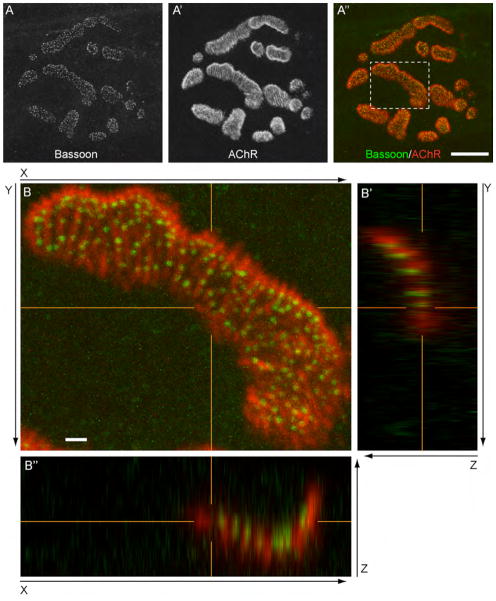
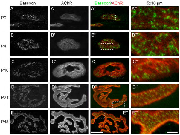
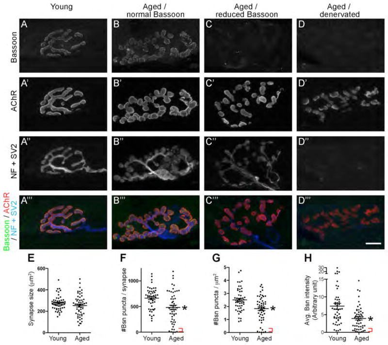
Presynaptic active zones are essential structures for synaptic vesicle release, but the developmental regulation of their number and maintenance during aging at mammalian neuromuscular junctions (NMJs) remains unknown. Here, we analyzed the distribution of active zones in developing, mature, and aged mouse NMJs by immunohistochemical detection of the active zone-specific protein Bassoon. Bassoon is a cytosolic scaffolding protein essential for the active zone assembly in ribbon synapses and some brain synapses. Bassoon staining showed a punctate pattern in nerve terminals and axons at the nascent NMJ on embryonic days 16.5-18.5. Three-dimensional reconstruction of NMJs revealed that the majority of Bassoon puncta within an NMJ were attached to the presynaptic membrane from postnatal day 0 to adulthood, and colocalized with another active zone protein, Piccolo. During postnatal development, the number of Bassoon puncta increased as the size of the synapses increased. Importantly, the density of Bassoon puncta remained relatively constant from postnatal day 0 to 54 at 2.3 puncta/μm(2) , while the synapse size increased 3.3-fold. However, Bassoon puncta density and signal intensity were significantly attenuated at the NMJs of 27-month-old aged mice. These results suggest that synapses maintain the density of synaptic vesicle release sites while the synapse size changes, but this density becomes impaired during aging.


Conflict of interest statement

The authors have no competing interests to declare.

Figures

Figure 1
Figure 1
Active zone protein Bassoon localizes at NMJ active zones. A - A″: The active zone-specific protein Bassoon forms puncta that are aligned with postsynaptic junctional folds in adult NMJs. Bassoon and acetylcholine receptors (AChRs) were visualized by fluorescent immunohistochemistry using an anti-Bassoon antibody (green) and Alexa Fluor 594-labeled α-bungarotoxin (red) in the sternomastoid muscle of P36 mice. Many Bassoon puncta were localized on top of the postsynaptic folds, which were visualized as bright lines of α-bungarotoxin staining. B - B″: The orthogonal views of the dotted area in the NMJ shown in (A″). The presynaptic terminal was placed toward the top of Z-axis, and the muscle fiber was placed toward the bottom. The panel B shows the XY-view of a maximal projected image. Panels B′ and B″ show YZ- and XZ-views of orthogonal single optical planes at the positions indicated by the orange lines in the panel B, XY-view. The orange lines in each image indicate the matching X, Y, and Z coordinates, respectively. The anti-Bassoon antibody (green) and Alexa Fluor 594-labeled α-bungarotoxin (red) revealed that all Bassoon puncta are attached to the α-bungarotoxin signal on the nerve side in the XZ- and YZ-orthogonal views, suggesting their anchoring to the presynaptic membrane. In the mature NMJ, the nerve terminal sinks into the muscle fiber and forms the primary gutter (Desaki and Uehara, 1987), which is shown as the U-shaped bungarotoxin staining in the XZ- and YZ-views. The maximal projected confocal images and the orthogonal views are shown without level adjustment. A magenta-green version of this figure is supplied as Supplementary Figure 1. Scale bars: (A – A″) 10 μm, (B – B″) 1 μm.
Figure 2
Figure 2
Bassoon and Piccolo puncta are anchored to the presynaptic membrane in adult NMJs. AA‴: The active zone-specific protein Piccolo (A′, red in A‴) showed a punctate staining pattern and overlapped with Bassoon staining (A, green in A‴) at a P36 NMJ. Many overlapping puncta were localized on top of the bright lines of Alexa Fluor 647-labeled α-bungarotoxin (A″, blue in A‴). B - B″: The orthogonal views of the NMJ shown in (A‴) with the same layout as the orthogonal views in Fig. 1B. Many overlapping puncta were attached to the α-bungarotoxin signal on the nerve side. Scale bars: 1 μm.
Figure 3
Figure 3
Bassoon is distributed as puncta in the nascent NMJs. AB″″: The active zone-specific protein Bassoon (A, A′, green in A‴, A″″, B‴, B″″) and AChRs (A″, red in A‴, A″″, B‴, B″″) were detected at E16.5 (A – A″″) and E18.5 (B – B″″), as described in Fig. 1. Arrowheads in panels A and B indicate Bassoon staining outside of the NMJs in a string-like pattern, suggesting axonal staining. The panels A′ and B′ show highly magnified regions indicated by the arrowheads in A and B. The panels A″″ and B″″ show a highly magnified 5 x 10 μm region indicated by the white dotted box in A‴ and B‴. C – C″: The orthogonal views of a P0 NMJ with the same layout as the orthogonal views in Fig 1B. The presynaptic terminal was placed toward the top of the Z-axis, and the muscle fiber was placed toward the bottom. The anti-Bassoon antibody (green) and Alexa Fluor 594-labeled α-bungarotoxin (red) revealed that most Bassoon puncta are attached to the bungarotoxin signal on the nerve side (white arrowheads), indicating that most Bassoon proteins are anchored to the presynaptic membrane. One Bassoon punctum was detected separate from the bungarotoxin-labeled endplate in YZ- and XZ-orthogonal views (white arrow in panels B′ and B″), suggesting that a small portion of the Bassoon puncta (0.6%, see main text) were floating in the presynaptic terminal at P0. The nascent NMJ lay relatively flat on the muscle fiber compared to the adult NMJs (Fig. 4B), which is shown by the flat α-bungarotoxin signal in the XZ-view (C″) and the A-shape in the YZ-view (C′). D: A representative transmission electron micrograph of a NMJ at E18.5. Active zones (green arrowheads) and a nascent postsynaptic junctional fold (white arrow) were observed. This in vivo data showed one electron-dense material that was not anchored to the presynaptic membrane and was floating in the nerve terminal (green arrow), which resembled the floating Bassoon puncta detected in (C). A magenta-green version of this figure is supplied as Supplementary Figure 2. Scale bars: (A, A″, A‴, B, B″, B‴) 10 μm, (A′, A″″, B′, B″″) 2 μm, (C – C″) 5 μm, (D) 500 nm.
Figure 4
Figure 4
Most Bassoon puncta were anchored to the presynaptic membrane at NMJs. A – A″: The orthogonal views of P0 NMJs are shown with the same layout as in Fig. 1B. The presynaptic terminal was placed toward the top of Z-axis, and the muscle fiber was placed toward the bottom. The anti-Bassoon antibody (green) and Alexa Fluor 594-labeled α-bungarotoxin (red) revealed that >99% of Bassoon puncta are attached to the bungarotoxin signal on the nerve side (white arrowheads), indicating that the majority of Bassoon proteins are anchored to the presynaptic membrane. The nascent NMJ lay flat on the muscle fiber, which is shown by the flat α-bungarotoxin signal in the YZ- and XZ-views (A′ and A″). B – B″: In the P48 NMJ, all Bassoon puncta are attached to the bungarotoxin signal on the nerve side (white arrowheads). The primary gutter is shown as the U-shaped bungarotoxin staining in the YZ- and XZ-views (B′ and B″). A magenta-green version of this figure is supplied as Supplementary Figure 3. Scale bars: 5 μm.
Figure 5
Figure 5
A-B′: Representative transmission electron micrographs showing active zones and postsynaptic junctional folds at the NMJs of sternocleidomastoid muscles at E18.5 (A) and P21 (B). Higher magnification images of A and B are shown in A′ and B′. Arrowheads indicate electron-dense material at the active zones that are aligned (black) or not aligned (white) with the junctional folds. The white arrows in panels A and A′ indicate the nascent junctional folds at E18.5. C: The quantification of the alignment between active zones and postsynaptic junctional folds by electron microscopy revealed an increase in the alignment during early postnatal development. The quantifications are from a total of 35 to 119 NMJ profiles per age. Analysis using a one-way ANOVA revealed that the increment between P4 and 2-week (wk)-old mice was significant (P < 0.05), but the increments between other immediately adjacent ages were not significant. Scale bars: (A – B′) 500 nm.
Figure 6
Figure 6
The postnatal development of the Bassoon puncta distribution at NMJs. The active zone-specific protein Bassoon (A, B, C, D, E; green in the right two columns) and AChRs (A′, B′, C′, D′, E′; red in the right two columns) were detected from P0 (A – A‴), P4 (B – B‴), P10 (C – C‴), P21 (D – D‴), and P48 (E – E‴), as described in Fig. 1. The three left panels show the entire NMJs. The rightmost panels (A‴ – E‴) show a highly magnified 5 x 10 μm region of the NMJs (white dotted boxes in A″ – E″). A magenta-green version of this figure is supplied as Supplementary Figure 4. Scale bars: (left three columns) 10 μm, (the most right column) 2 μm.
Figure 7
Figure 7
The constant density of the Bassoon puncta at postnatal NMJs. A: The synapse size showed constant growth from the embryonic to adult stages, which was determined by the α-bungarotoxin stained area (P0: 89.7 ± 5.5; P4: 125.9 ± 7.8; P10: 179.2 ± 7.3; P21: 217.9 ± 9.5; P48 –54: 295.2 ± 17.6 μm2). B: The number of Bassoon puncta increased as the synapse size increased. The average number of Bassoon puncta in a synapse constantly increased from P0 to P54 (P0: 183.0 ± 12.7; P4: 304.3 ± 25.4; P10: 365.7 ± 24.8; P21: 553.2 ± 24.5; P48–54: 780.2 ± 45.4). C: The density of the Bassoon puncta stayed at a relatively constant level (2.3 puncta/μm2) during the postnatal development of the NMJs (P0: 2.0 ± 0.09; P4: 2.3 ± 0.1; P10: 1.9 ± 0.1; P21: 2.5 ± 0.1; P54: 2.6 ± 0.1 puncta/μm2). These quantifications were from 31–39 NMJs from 3 mice at each stage. Asterisks indicate significance by one-way ANOVA analysis (P < 0.05). (A) The synapse size at P10 and P48–54 was significantly larger than the immediately adjacent younger age groups. (B) The Bassoon puncta number per synapse was significantly larger than the immediately adjacent younger age groups at P4, P21, and P54. (C) Significant differences were detected between P10 and P21.
Figure 8
Figure 8
The active zone protein Bassoon was reduced in the NMJs of aged mice. AD‴: The sternomastoid muscles of aged mice (27-month-old, B – D‴) and young adult mice (1-month-old, A – A‴) were stained using an anti-Bassoon antibody (active zone, A, B, C, D; green in A‴, B‴, C‴, D‴), Alexa Fluor 594-labeled α-bungarotoxin (AChR, A′, B′, C′, D′; red in A‴, B‴, C‴, D‴), and anti-neurofilament plus anti-SV2 antibodies (nerve morphology, A″, B″, C″, D″; blue in A‴, B‴, C‴, D‴). Innervation was confirmed by the nerve morphology visualized using anti-neurofilament plus anti-SV2 antibodies. Panels D – D‴ show a denervated aged NMJ. Bassoon puncta were significantly attenuated in some aged NMJs (C – C‴), while other aged NMJs retained near normal levels of Bassoon staining (B – B‴). Innervated NMJs of aged mice had a similar synapse size (E: young adult mice, 276.2 ± 9.8 μm2; aged mice, 253.3 ± 13.9 μm2; P = 0.19) as the young adult mice. However, the NMJs of aged mice had a decreased number of Bassoon puncta per synapse (F: young adult mice, 664.3 ± 29.9; aged mice, 482.3 ± 43.3; P = 0.0009), a reduced density of Bassoon puncta (G: young adult mice, 2.5 ± 0.1 puncta/μm2; aged mice, 1.8 ± 0.1 puncta/μm2; P = 0.0005), and attenuated Bassoon average signal intensity (H, in arbitrary intensity units: young adult mice, 7.4 ± 0.8; aged mice, 3.9 ± 0.5; P = 0.0003). The red brackets in F – H indicate a subgroup of the NMJs of aged mice with a minuscule level of Bassoon signal at the synapses. The quantifications (E – H) are from a total of 52 NMJs from four aged mice and 49 NMJs from four young adult mice. Asterisks indicate significant differences by unpaired t-test. Scale bar: 10 μm.

Similar articles

Cited by

References

    1. Ahmari SE, Buchanan J, Smith SJ. Assembly of presynaptic active zones from cytoplasmic transport packets. Nat Neurosci. 2000;3(5):445–451. - PubMed
    1. Alshuaib WB, Fahim MA. Depolarization reverses age-related decrease of spontaneous transmitter release. J Appl Physiol. 1991;70(5):2066–2071. - PubMed
    1. Balice-Gordon R, Lichtman J. In vivo observations of pre- and postsynaptic changes during the transition from multiple to single innervation at developing neuromuscular junctions. The Journal of Neuroscience. 1993;13(2):834–855. - PMC - PubMed
    1. Balice-Gordon RJ. Age-related changes in neuromuscular innervation. Muscle Nerve Suppl. 1997;5:S83–87. - PubMed
    1. Banker BQ, Kelly SS, Robbins N. Neuromuscular transmission and correlative morphology in young and old mice. The Journal of physiology. 1983;339:355–377. - PMC - PubMed

Publication types