Comparative Study
. 2012 Apr 1;520(5):1047-61.
doi: 10.1002/cne.22769.

Pre-Bötzinger complex receives glutamatergic innervation from galaninergic and other retrotrapezoid nucleus neurons

Affiliations
Comparative Study

Pre-Bötzinger complex receives glutamatergic innervation from galaninergic and other retrotrapezoid nucleus neurons

Genrieta Bochorishvili et al. J Comp Neurol. .

Abstract

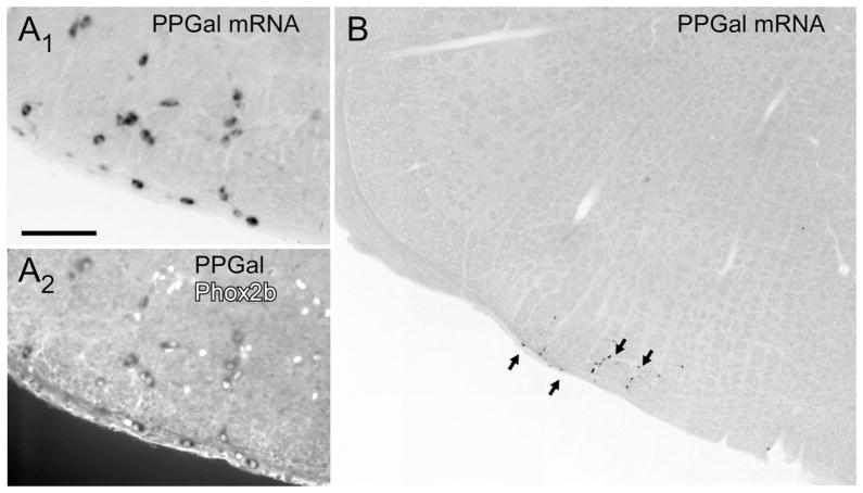
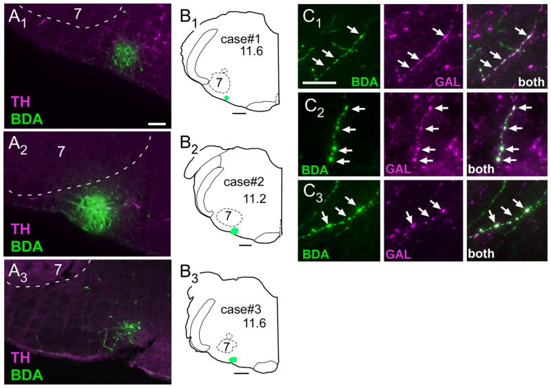
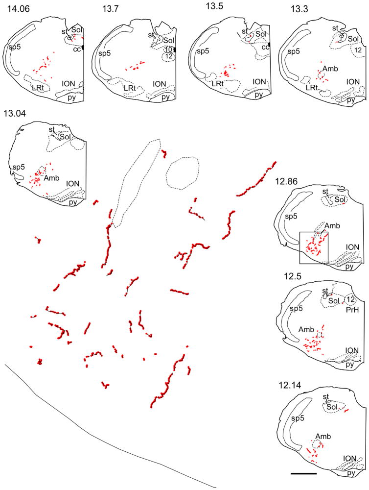
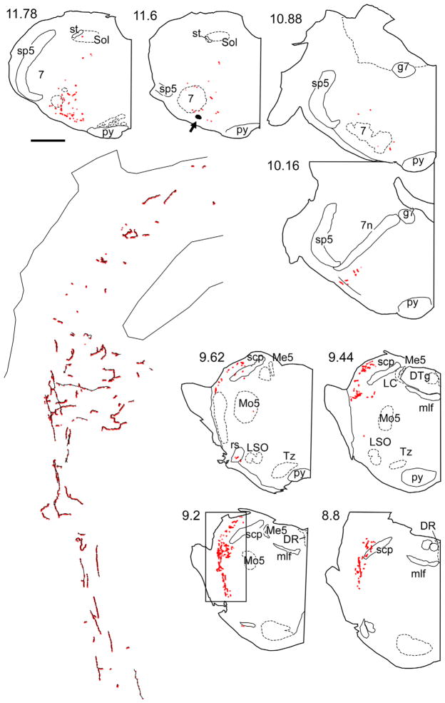
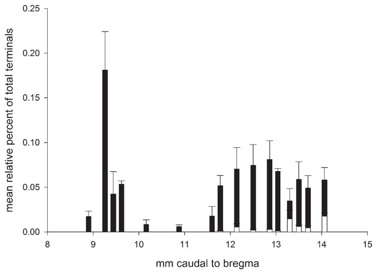
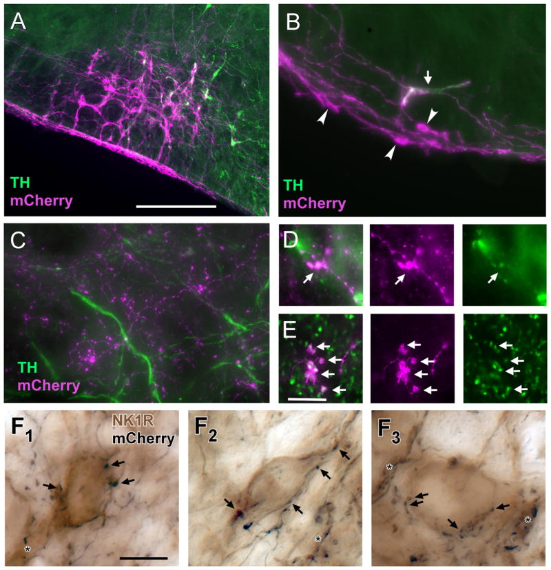
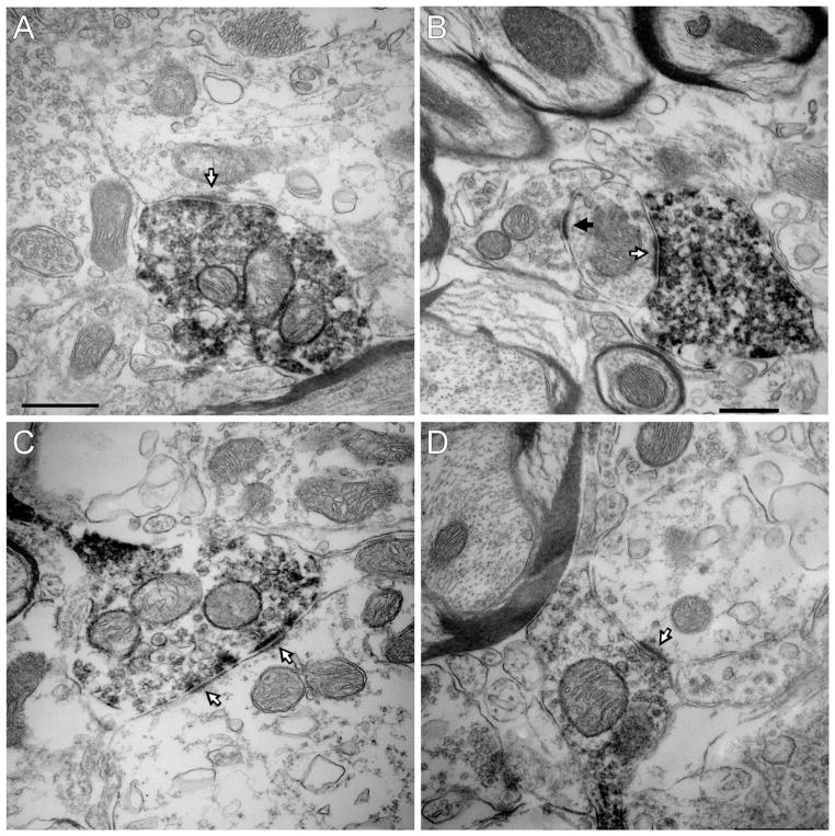
The retrotrapezoid nucleus (RTN) contains CO(2) -responsive neurons that regulate breathing frequency and amplitude. These neurons (RTN-Phox2b neurons) contain the transcription factor Phox2b, vesicular glutamate transporter 2 (VGLUT2) mRNA, and a subset contains preprogalanin mRNA. We wished to determine whether the terminals of RTN-Phox2b neurons contain galanin and VGLUT2 proteins, to identify the specific projections of the galaninergic subset, to test whether RTN-Phox2b neurons contact neurons in the pre-Bötzinger complex, and to identify the ultrastructure of these synapses. The axonal projections of RTN-Phox2b neurons were traced by using biotinylated dextran amine (BDA), and many BDA-ir boutons were found to contain galanin immunoreactivity. RTN galaninergic neurons had ipsilateral projections that were identical with those of this nucleus at large: the ventral respiratory column, the caudolateral nucleus of the solitary tract, and the pontine Kölliker-Fuse, intertrigeminal region, and lateral parabrachial nucleus. For ultrastructural studies, RTN-Phox2b neurons (galaninergic and others) were transfected with a lentiviral vector that expresses mCherry almost exclusively in Phox2b-ir neurons. After spinal cord injections of a catecholamine neuron-selective toxin, there was a depletion of C1 neurons in the RTN area; thus it was determined that the mCherry-positive terminals located in the pre-Bötzinger complex originated almost exclusively from the RTN-Phox2b (non-C1) neurons. These terminals were generally VGLUT2-immunoreactive and formed numerous close appositions with neurokinin-1 receptor-ir pre-Bötzinger complex neurons. Their boutons (n = 48) formed asymmetric synapses filled with small clear vesicles. In summary, RTN-Phox2b neurons, including the galaninergic subset, selectively innervate the respiratory pattern generator plus a portion of the dorsolateral pons. RTN-Phox2b neurons establish classic excitatory glutamatergic synapses with pre-Bötzinger complex neurons presumed to generate the respiratory rhythm.


Figures

Figure 1
Figure 1
Preprogalanin (PPGal) mRNA in the retrotrapezoid nucleus. A1: PPGal mRNA-containing neurons (brightfield illumination) with mRNA revealed by nonradioactive digoxigenin immunohistochemistry using alkaline phosphatase and nitroblue tetrazolium/5-bromo-4-chloro-3-indolyl-phosphate as substrate (see Stornetta et al., 2009 for details). A2: Same field photographed under fluorescent light to reveal the Phox2b-immnoreactive (ir) nuclei. Every PPGal-mRNA-positive neuron contains a Phox2b-ir nucleus. B: Photomontage illustrating the isolation of the cluster of PPGal-mRNA-positive retrotrapezoid nucleus (RTN) neurons. Scale bar in A1 = 100 μm for A1, A2; 330 μm for B.
Figure 2
Figure 2
BDA injection sites and anterograde labeling of galaninergic axonal processes. A1–A3: Injection sites of biotinylated dextran amine (BDA) in three rats. The iontophoretic injections of BDA (revealed with Alexa-488, green fluorescence) were deliberately targeted below the caudal edge of the facial motor nucleus where the highest density of RTN Phox2b neurons reside. This region sits at the lateral edge of the column of C1 adrenergic neurons (identified by immunohistochemical detection of tyrosine-hydroxylase [TH] with Cy-3, in magenta). B1–B3: Computer-generated drawings of the location in coronal sections of the three BDA injection sites shown in A. The numbers below the case numbers refer to mm behind bregma after the atlas of Paxinos and Watson (1998). C1–C3: Examples of galanin-ir terminals (shown in magenta) containing BDA (BDA alone is green, double-labeled terminals appear white) within the pre-Bötzinger complex (C1), Kölliker-Fuse nucleus (C2), and lateral parabrachial nucleus (C3). Scale bar = 100 μm in A1 (applies to A1–A3); 500 μm in B1–B3; 10 μm in C1 (applies to C1–C3).
Figure 3
Figure 3
Representative example of CNS projections of the retrotrapezoid galaninergic neurons. The axonal varicosities that were immunoreactive for BDA and galanin are selectively represented as red dots on these computer-assisted drawings of coronal hemisections. The numbers to the upper left of each coronal hemisection drawing indicate the location of the section as the approximate number of mm caudal to bregma after the atlas of Paxinos and Watson (1998). The area in the rectangular box in section 12.86 corresponds to the pre-Bötzinger complex, and an enlargement showing fibers in black and boutons in red is shown in the middle. In the continuation panel, the area in the rectangular box in section 9.2 corresponds to the lateral parabrachial nucleus/Kölliker Fuse area, and an enlargement showing fibers in black and boutons in red is shown on the left side on the continuation panel. The arrow at level 11.6 mm caudal to Bregma points to the BDA injection site. Abbreviations: 7, facial nucleus; 12, hypoglossal nucleus; 7n, facial nucleus root; Amb, ambiguus nucleus; cc, central canal; DR, dorsal raphe nucleus; DTg, dorsal tegmental nucleus; g7, genu of facial nucleus; ION, inferior olivary nucleus; LRt, lateral reticular nucleus; LSO, lateral superior olive; Me5, mesencephalic 5 nucleus; mlf, medial longitudinal fasciculus; Mo5, motor trigeminal nucleus; py, pyramidal tract; rs, rubrospinal tract; scp, superior cerebellar peduncle; Sol, nucleus of the solitary tract; Sp5, spinal trigeminal nucleus; st, solitary tract; Tz, nucleus of trapezoid body. Scale bar = 1 mm for all smaller drawings, 0.09 mm for the enlargement representing the pre-Bötzinger complex, and 0.154 mm for the enlargement representing the parabrachial/Kölliker Fuse area. [Color figure can be viewed in the online issue, which is available at wileyonlinelibrary.com.]
Figure 3
Figure 3
Representative example of CNS projections of the retrotrapezoid galaninergic neurons. The axonal varicosities that were immunoreactive for BDA and galanin are selectively represented as red dots on these computer-assisted drawings of coronal hemisections. The numbers to the upper left of each coronal hemisection drawing indicate the location of the section as the approximate number of mm caudal to bregma after the atlas of Paxinos and Watson (1998). The area in the rectangular box in section 12.86 corresponds to the pre-Bötzinger complex, and an enlargement showing fibers in black and boutons in red is shown in the middle. In the continuation panel, the area in the rectangular box in section 9.2 corresponds to the lateral parabrachial nucleus/Kölliker Fuse area, and an enlargement showing fibers in black and boutons in red is shown on the left side on the continuation panel. The arrow at level 11.6 mm caudal to Bregma points to the BDA injection site. Abbreviations: 7, facial nucleus; 12, hypoglossal nucleus; 7n, facial nucleus root; Amb, ambiguus nucleus; cc, central canal; DR, dorsal raphe nucleus; DTg, dorsal tegmental nucleus; g7, genu of facial nucleus; ION, inferior olivary nucleus; LRt, lateral reticular nucleus; LSO, lateral superior olive; Me5, mesencephalic 5 nucleus; mlf, medial longitudinal fasciculus; Mo5, motor trigeminal nucleus; py, pyramidal tract; rs, rubrospinal tract; scp, superior cerebellar peduncle; Sol, nucleus of the solitary tract; Sp5, spinal trigeminal nucleus; st, solitary tract; Tz, nucleus of trapezoid body. Scale bar = 1 mm for all smaller drawings, 0.09 mm for the enlargement representing the pre-Bötzinger complex, and 0.154 mm for the enlargement representing the parabrachial/Kölliker Fuse area. [Color figure can be viewed in the online issue, which is available at wileyonlinelibrary.com.]
Figure 4
Figure 4
Rostrocaudal distribution of boutons containing BDA and galanin immunoreactivity. Frequency distribution histogram of double-labeled boutons based on the three BDA cases shown in Figure 2. The boutons located at levels between 8 and 10 mm behind bregma were all within the dorsolateral pons (closed bars). The boutons located caudal to the region at 11 mm behind bregma were in two regions (ventrolateral medulla and nucleus of the solitary tract) and are represented separately (closed and open bars, respectively).
Figure 5
Figure 5
Terminals of RTN-Phox2b neurons make close appositions with NK1R-ir neurons in the pre-Bötzinger complex. A: Example of mCherry-ir neurons within the RTN region 3 weeks after injection of PRSX8 ChR2-mCherry lentivirus into rats pretreated with anti-DβH-saporin to reduce the number of spinally projecting TH neurons. The vast majority of the transfected neurons are not TH-ir (TH immunofluorescence in green; ChR2-expressing TH neurons in white). B: Higher magnification example showing a single TH-ir neuron (arrow) and multiple TH-negative neurons (arrowheads) that contain mCherry (magenta). The depicted region is below the facial motor nucleus. The TH-negative neurons are located within the marginal layer of the RTN. C: The mCherry-ir terminals (magenta) located within the pre-Bötzinger complex do not contain TH immunoreactivity (green). D: mCherry-ir terminals (magenta) located within the raphe pallidus are TH-ir (TH in green, double-labeled terminals appear white). E: A large fraction of the mCherry-ir (magenta) terminals located within the Pre-Bötzinger complex are positive for VGLUT2 (green, double-labeled terminals appear white). F1–F3: Examples of close appositions between mCherry-ir varicosities (nickel-enhanced DAB reaction, black) and NK1R-ir somata or dendrites within the pre-Bötzinger complex (light brown). Some of the close appositions (putative synapses) are indicated by arrows. Close appositions on dendrites are indicated by the asterisks. Scale bar = 250 μm in A (applies to A–C); 10 μm in E (applies to D, E); 10 μm in F1 (applies to F1–F3).
Figure 6
Figure 6
Ultrastructure of the synaptic contacts between RTN-Phox2b neurons and pre-Bötzinger complex neurons. A–D: The mCherry-ir terminals of the RTN-Phox2b neurons contain intracellular and plasma membrane-associated electron-dense immunoperoxidase reaction product. These terminals make asymmetric synaptic contacts with unlabeled dendrites. The white arrows identify the thick postsynaptic material characteristic of excitatory glutamatergic synapses. The black arrow points to an asymmetric contact between an unlabeled terminal and a dendrite. Scale bar = 0.5 μm in A (applies to A, C, D); 0.5 μm in B.

References

    1. Abbott SB, Burke PG, Pilowsky PM. Galanin microinjection into the PreBötzinger or the Bötzinger complex terminates central inspiratory activity and reduces responses to hypoxia and hypercapnia in rat. Respir Physiol Neurobiol. 2009a;167:299–306. - PubMed
    1. Abbott SB, Stornetta RL, Socolovsky CS, West GH, Guyenet PG. Photostimulation of channelrhodopsin-2 expressing ventrolateral medullary neurons increases sympathetic nerve activity and blood pressure in rats. J Physiol. 2009b;587:5613–5631. - PMC - PubMed
    1. Abbott SBG, Stornetta RL, Fortuna MG, Depuy SD, West GH, Harris TE, Guyenet PG. Photostimulation of retrotrapezoid nucleus Phox2b-expressing neurons in vivo produces long-lasting activation of breathing in rats. J Neurosci. 2009c;29:5806–5819. - PMC - PubMed
    1. Alheid GF, Gray PA, Jiang MC, Feldman JL, McCrimmon DR. Parvalbumin in respiratory neurons of the ventrolateral medulla of the adult rat. J Neurocytol. 2002;31:693–717. - PubMed
    1. Boscan P, Dutschmann M, Herbert H, Paton JF. Neurokininergic mechanism within the lateral crescent nucleus of the parabrachial complex participates in the heart-rate response to nociception. J Neurosci. 2005;25:1412–1420. - PMC - PubMed

Publication types