. 2012 Mar 1;520(4):756-69.
doi: 10.1002/cne.22773.

Cone outer segment extracellular matrix as binding domain for interphotoreceptor retinoid-binding protein

Affiliations

Cone outer segment extracellular matrix as binding domain for interphotoreceptor retinoid-binding protein

Mary Alice Garlipp et al. J Comp Neurol. .

Abstract

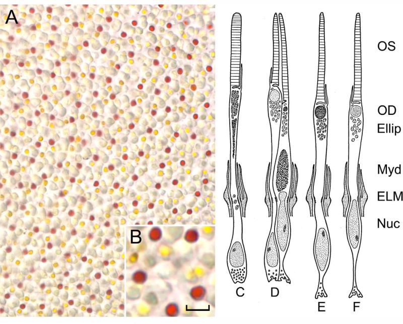
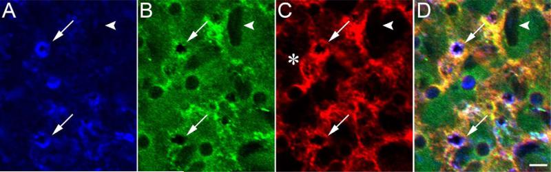
Cones are critically dependent on interphotoreceptor retinoid-binding protein (IRBP) for retinoid delivery in the visual cycle. Cone-dominant vertebrates offer an opportunity to uncover the molecular basis of IRBP's role in this process. Here, we explore the association of IRBP with the interphotoreceptor matrix (IPM) of cones vs. rods in cone dominant retinas from chicken (Gallus domesticus), turkey (Meleagris gallopavo), and pig (Sus scrofa). Retinas were detached and fixed directly or washed in saline prior to fixation. Disassociated photoreceptors with adherent matrix were also prepared. Under 2 mM CaCl(2) , insoluble matrix was delaminated from saline washed retinas. The distribution of IRBP, as well as glycans binding peanut agglutinin (cone matrix) and wheat germ agglutinin (rod/cone matrix), was defined by confocal microscopy. Retina flat mounts showed IRBP diffusely distributed in an interconnecting, lattice-like pattern throughout the entire matrix. Saline wash replaced this pattern with fluorescent annuli surrounding individual cone outer segments. In isolated cones and matrix sheets, IRBP colocalized with the peanut agglutinin binding matrix glycans. Our results reveal a wash-resistant association of IRBP with a matrix domain immediately surrounding cone outer segments. The cone matrix sheath may be responsible for IRBP-mediated cone targeting of 11-cis retinoids.


Figures

Figure 1
Figure 1
Chicken photoreceptor subtypes. A: The retina flat mount shows the cone cell mosaic with its various colored oil droplets (inset in B). There are four visual cells: rod (C; 13%), no oil droplet; double cone (D; 36%), consisting of larger principal cone with golden-yellow oil droplet, and smaller accessory cone with green-yellow oil droplet; single cone type I (E; 25%), dark red oil droplet; single cone type II (F; 11%), light colored oil droplet. OS, outer segment; OD, oil droplet, Ellip, ellipsoid; Myd, myoid; ELM, external limiting membrane; Nuc, nucleus. Diagram adapted from Morris and Shorey (J. Comp. Neurol. 129:313–340, 1997) with permission from John Wiley & Sons, Inc. Scale bar = 10 μm in A, 5 μm in B.
Figure 2
Figure 2
Effect of saline wash on IRBP distribution. Unwashed chicken retinas were detached directly under 4% paraformaldehyde (A–D), or washed retinas were detached under Ringer's saline (E–H). Retina flat mounts were probed with mAb F7 anti-IRBP, followed by goat anti-mouse IgG-647. A,E: Differential interference contrast microscopy (DIC). B,F: Confocal IRBP immunofluorescence (633 nm). C,G: Fluorescence merged with DIC. D,H: Same tissue following removal from coverslipped slide and reoriented for cross-sections (fuorescence merged with DIC). Oil droplet (arrow), outer segments (arrowhead). A magenta–green copy of this figure is available as Supporting Information Figure 2. Scale bar = 10 μm.
Figure 3
Figure 3
IRBP distribution in saline washed chicken retina. Retinas were washed three times, flat mounted, and probed with mAb F7 anti-IRBP, followed by goat anti-mouse IgG-647. Overview of merged confocal fluorescence images at 633 nm (red, IRBP) and 488 nm (green, oil droplet autofluorescence). A: En face orientation of cone array of IRBP indirect immunofluorescence (arrow, double cone). B: En face orientation incubated without primary antibody. C: Oblique section showing inner segment (IS) and outer segment (OS) zones. Arrows, distal cone outer segments tips. D: Enlargement of marked double-cone region in A. Arrowheads, ring-like association of IRBP with cone outer segments; asterisk, region between IRBP cone-associated domains. A magenta–green copy of this figure is available as Supporting Information Figure 3. Scale bar = 3.3 μm in D; 10 μm for A–C.
Figure 4
Figure 4
Localization of IRBP and PNA-binding glycans in chicken (A–C) and turkey (D–F) retinas. Isolated retinas were probed with mAb F7 anti-IRBP, followed by goat anti-mouse IgG-647 (A,D) and PNA-488 (B,E). C,F: Merged fluorescence. Omission of the primary antibody or PNA-488 showed no fluorescence (data not illustrated). Arrows correspond to cone matrix sheath magnified in insets. A magenta–green copy of this figure is available as Supporting Information Figure 4. Scale bar = 2 μm in insets; 10 μm for A–F.
Figure 5
Figure 5
Localization of IRBP and PNA-binding glycans in the pig retina. Isolated retinas were probed with mAb F7 anti-IRBP, followed by goat anti-mouse IgG-647 (A) or PNA-488 (B). Merged fluorescence is shown in C. IRBP and PNA-binding glycans show strong colocalization (arrow). A magenta–green copy of this figure is available as Supporting Information Figure 5. Scale bar = 10 μm.
Figure 6
Figure 6
Isolated turkey cones probed with PNA-488 (A–C) or WGA-647 (D–F). Washed retinas were incubated with either lectin prior to gentle trituration to generate CIS/COS. A,D: Retained cone matrix sheath (arrowheads) is detectable around the outer and inner segments by DIC. B,E: Confocal immunofluorescence of PNA-488 and WGA-647 fluorescence, respectively. C,F: Merged fluorescence and DIC. OS, outer segment; OD, oil droplets; Ellip, ellipsoid; Myd, myoid; arrow, PNA-positive cone matrix sheath. A magenta–green copy of this figure is available as Supporting Information Figure 6. Scale bar = 5 μm.
Figure 7
Figure 7
Localization of IRBP and PNA-binding glycans in dissociated turkey photoreceptors. Washed retinas were incubated with mAb F7 anti-IRBP followed by goat anti-mouse IgG-647. The tissue was then probed with PNA-488 and gently triturated to generate photoreceptor clusters or CIS/COS. A–C show a single cone (outer segment marked with arrow) and nearby rod outer segment (arrowhead); D–F show a double cone. A,D: Cone matrix sheath labeling with PNA-488. B,E: IRBP localization within the cone matrix sheath. C,F: Merged corresponding fluorescence and DIC images. Note that IRBP colocalization within the cone matrix sheath is restricted to an outer segment subdomain (yellow merged fluorescence). Ellip, ellipsoid; OD, oil droplet. A magenta–green copy of this figure is available as Supporting Information Figure 7. Scale bar = 8.7 μm in F (applies to D–F); 5 μm for A–C.
Figure 8
Figure 8
Localization of IRBP and PNA-binding glycans in isolated chicken IPM. Matrix sheets were delaminated from retinas in 2 mM CaCl2 and mounted onto subbed slides. A: Indirect IRBP immunofluorescence using mAb F7 anti-IRBP detected with goat anti-mouse IgG-647. B: Distribution of PNA-488-binding glycans. C: Merged fluorescence. Asterisk, matrix appearing to bridge between cone matrix sheath structures; arrows, fluorescence colocalization in cone matrix sheaths. A magenta–green copy of this figure is available as Supporting Information Figure 8. Scale bar = 10 μm.
Figure 9
Figure 9
Localization of IRBP and PNA- and WGA-binding glycans within isolated turkey IPM. The matrix sheet was delaminated from a freshly isolated retina in 2 mM CaCl2 and probed with MAb F7 anti-IRBP followed by goat-anti mouse IgG-647 (A), PNA-488 (B), and WGA-555 (C). D: Merged IRBP, PNA, and WGA fluorescence. IRBP immunofluorescence is shown in blue because three fluorophores are used (647-blue, 633 nm; 488-green, 488 nm; 555-red, 561 nm). Asterisk, WGA-positive matrix appearing to bridge nearby cone sheaths; arrows, colocalization of IRBP and PNA/WGA-binding glycans; arrowheads, rod-associated matrix. A magenta–green copy of this figure is available as Supporting Information Figure 9. Scale bar = 5 μm.

References

    1. Adler AJ, Evans CD. Rapid isolation of bovine interphotoreceptor retinol-binding protein. Biochim Biophys Acta. 1983;761:217–222. - PubMed
    1. Adler AJ, Chader GJ, Wiggert B. Purification and assay of interphotoreceptor retinoid-binding protein from the eye. Methods Enzymol. 1990;189:213–223. - PubMed
    1. Anderson DH, Neitz J, Saari JC, Kaska DD, Fenwick J, Jacobs GH, Fisher SK. Retinoid-binding proteins in cone-dominant retinas. Invest Ophthalmol Vis Sci. 1986;27:1015–1026. - PubMed
    1. Biernbaum MS, Bownds MD. Frog rod outer segments with attached inner segment ellipsoids as an in vitro model for photoreceptors on the retina. J Gen Physiol. 1985;85:83–105. - PMC - PubMed
    1. Blanks JC, Johnson LV. Specific binding of peanut lectin to a class of retinal photoreceptor cells. A species comparison. Invest Ophthalmol Vis Sci. 1984;25:546–557. - PubMed

Publication types

LinkOut - more resources