. 2011 Sep 21:7:71.
doi: 10.1186/1744-8069-7-71.

Migraine attacks the Basal Ganglia

Affiliations

Migraine attacks the Basal Ganglia

Nasim Maleki et al. Mol Pain. .

Abstract

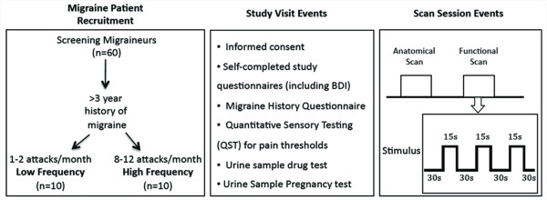
Background: With time, episodes of migraine headache afflict patients with increased frequency, longer duration and more intense pain. While episodic migraine may be defined as 1-14 attacks per month, there are no clear-cut phases defined, and those patients with low frequency may progress to high frequency episodic migraine and the latter may progress into chronic daily headache (> 15 attacks per month). The pathophysiology of this progression is completely unknown. Attempting to unravel this phenomenon, we used high field (human) brain imaging to compare functional responses, functional connectivity and brain morphology in patients whose migraine episodes did not progress (LF) to a matched (gender, age, age of onset and type of medication) group of patients whose migraine episodes progressed (HF).

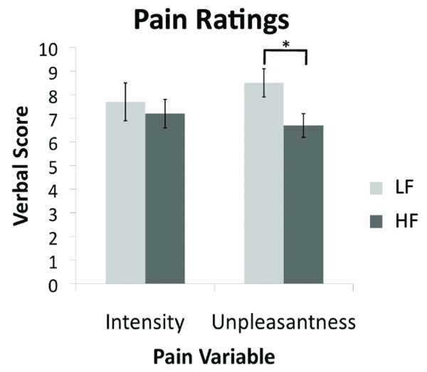
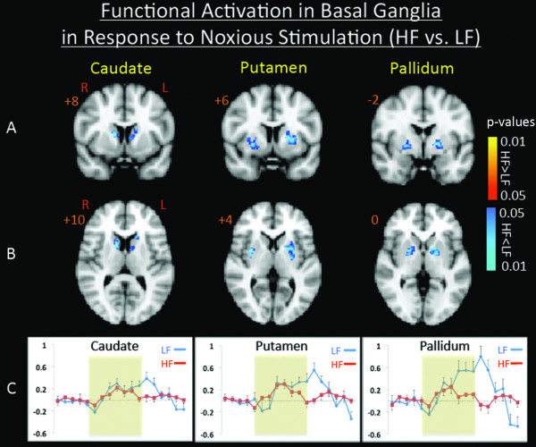
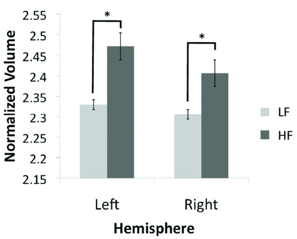
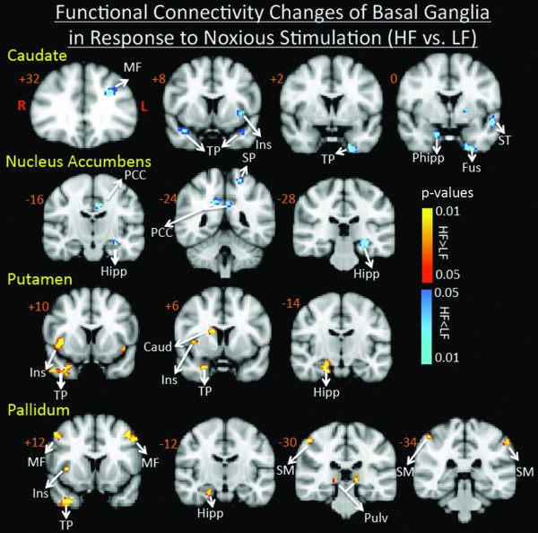
Results: In comparison to LF patients, responses to pain in HF patients were significantly lower in the caudate, putamen and pallidum. Paradoxically, associated with these lower responses in HF patients, gray matter volume of the right and left caudate nuclei were significantly larger than in the LF patients. Functional connectivity analysis revealed additional differences between the two groups in regard to response to pain.

Conclusions: Supported by current understanding of basal ganglia role in pain processing, the findings suggest a significant role of the basal ganglia in the pathophysiology of the episodic migraine.


Figures

Figure 1
Figure 1
Migraine Pain Intensity and Unpleasantness Ratings. There is a significant difference in the pain unpleasantness scores between the two cohorts (p < 0.028). The scores are based on a 0-10 subjective scale for migraine pain intensity and pain unpleasantness.
Figure 2
Figure 2
Contrast Maps for Painful Heat fMRI Activation. Contrast analysis of the HF vs. LF migraine group in response to the "pain threshold +1°C" stimuli revealed significant (p < 0.05, corrected) differences in the basal ganglia nuclei in coronal (A) and axial (B) views. In (C) Single trial averages for the response to painful heat stimulation in caudate, putamen, and pallidum are presented. Yellow represents the stimulus application period.
Figure 3
Figure 3
Volumetric Changes of the Caudate. The plots show the significant caudate volumetric differences in the high vs. low frequency migraine subjects (left: p < 0.025 and right: p < 0.006). The volumes have been normalized to the total intracranial volume to scale for the brain volume for each subject. Bar heights represent the mean value for each volumetric measurement. Error bars represent the 95% confidence interval of the mean. * denotes significance.
Figure 4
Figure 4
Functional Connectivity Contrast Maps of the Basal Ganglia Nuclei. Functional connectivity contrast maps of the basal ganglia nuclei during intermittent heat stimuli (pain threshold +1°C on hand) in high frequency migraine patients vs. low frequency migraine patients. PCC: Posterior Cingulate Cortex, SM: SupraMarginal, SF: Superior Frontal, ST: Superior Temporal, SP: Superior Parietal, Ins: Insula, Hipp: Hippocampus, PHipp: Parahippocampus, Fus: Fusiform, Thal: Thalamus, Pulv: Pulvinar, TP: Temporal Pole, MF: Middle Frontal.
Figure 5
Figure 5
Subject Recruitment and Experimental Approach. Based on the frequency of migraine attacks per month, Low frequency (LF) and High frequency (HF) episodic migraine patients were recruited to the study. During the study visit Quantitative Sensory Testing (QST) for pain threshold was performed for each subject and questionnaires were filled out. For each subject morphological (as part of the anatomical scan) and functional (evoked to thermal stimuli) images were acquired as shown in the diagram. Thermal stimuli paradigm is also shown in the diagram.

References

    1. Olesen J, Ashina M. Emerging migraine treatments and drug targets. Trends Pharmacol Sci. 2011;32:352–359. doi: 10.1016/j.tips.2011.02.016. - DOI - PubMed
    1. Bussone G. Pathophysiology of migraine. Neurol Sci. 2004;25( Suppl 3):S239–241. - PubMed
    1. May A. New insights into headache: an update on functional and structural imaging findings. Nat Rev Neurol. 2009;5:199–209. doi: 10.1038/nrneurol.2009.28. - DOI - PubMed
    1. Geuze E, Westenberg HG, Heinecke A, de Kloet CS, Goebel R, Vermetten E. Thinner prefrontal cortex in veterans with posttraumatic stress disorder. Neuroimage. 2008;41:675–681. doi: 10.1016/j.neuroimage.2008.03.007. - DOI - PubMed
    1. Bartolini M, Baruffaldi R, Paolino I, Silvestrini M. Cerebral blood flow changes in the different phases of migraine. Funct Neurol. 2005;20:209–211. - PubMed

Publication types