

A case of bilateral self-induced keratoconus in a patient with tourette syndrome associated with compulsive eye rubbing: case report
- PMID: 21936935
- PMCID: PMC3191478
- DOI: 10.1186/1471-2415-11-28
A case of bilateral self-induced keratoconus in a patient with tourette syndrome associated with compulsive eye rubbing: case report
Abstract
Background: Tourette syndrome is a neurologic disorder that is characterized by repetitive muscle contractions that produce stereotyped movements or sounds. Approximately 50% of individuals with TS also exhibit obsessive-compulsive behaviors including eye rubbing. We report a case of bilateral self-induced keratoconus in a patient with TS, associated with compulsive eye rubbing.
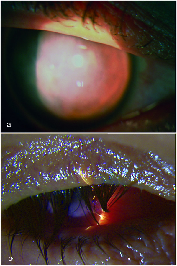
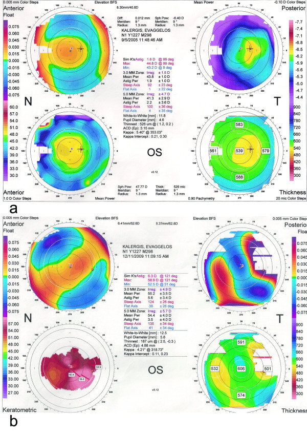
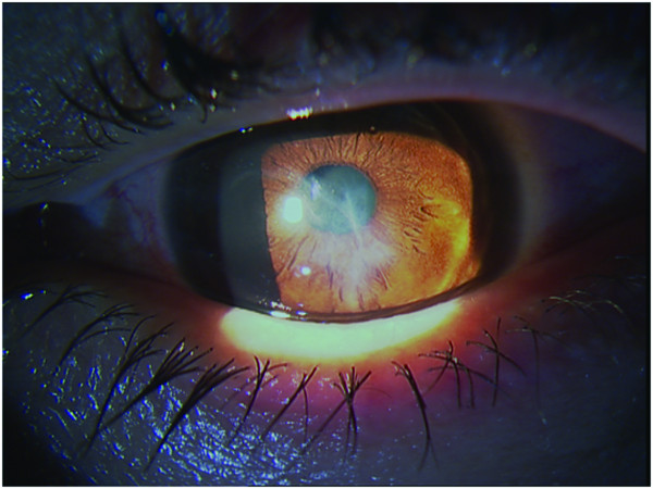
Case presentation: A 35-year-old man was first seen in our clinic as an outpatient due to rapid deterioration of vision in his right eye associated with pain and tearing, over a period of one month. Slit lamp biomicroscopy of the right eye showed a central stromal scar due to corneal hydrops. Clinical examination and corneal topography of the left eye were normal. Six months later the patient developed corneal hydrops of his left eye. During the following examinations his vision continued to deteriorate in both eyes, while a central stromal scar was forming in his left cornea. Four years after the initial examination the patient's visual acuity was no light perception in the right eye and counting fingers at 33 cm in the left eye. His right eye was phthisic.
Conclusions: Our patient developed a rapidly progressing bilateral corneal ectasia and phthisis of his right eye during a time period of 4 years. This unusual pattern suggests that the patient's compulsive behavior compromised both of his corneas and led to bilateral keratoconus.
Figures
References
-
- American Psychiatric Association. Diagnostic and Statistical Manual of Mental Disorders. American Psychiatric Association; 1994.
Publication types
MeSH terms
LinkOut - more resources
Full Text Sources
Medical
