Case Reports
. 2013 May 10;165(2):314-21.
doi: 10.1016/j.ijcard.2011.08.079. Epub 2011 Sep 19.

Novel MYH11 and ACTA2 mutations reveal a role for enhanced TGFβ signaling in FTAAD

Affiliations
Case Reports

Novel MYH11 and ACTA2 mutations reveal a role for enhanced TGFβ signaling in FTAAD

Marjolijn Renard et al. Int J Cardiol. .

Abstract

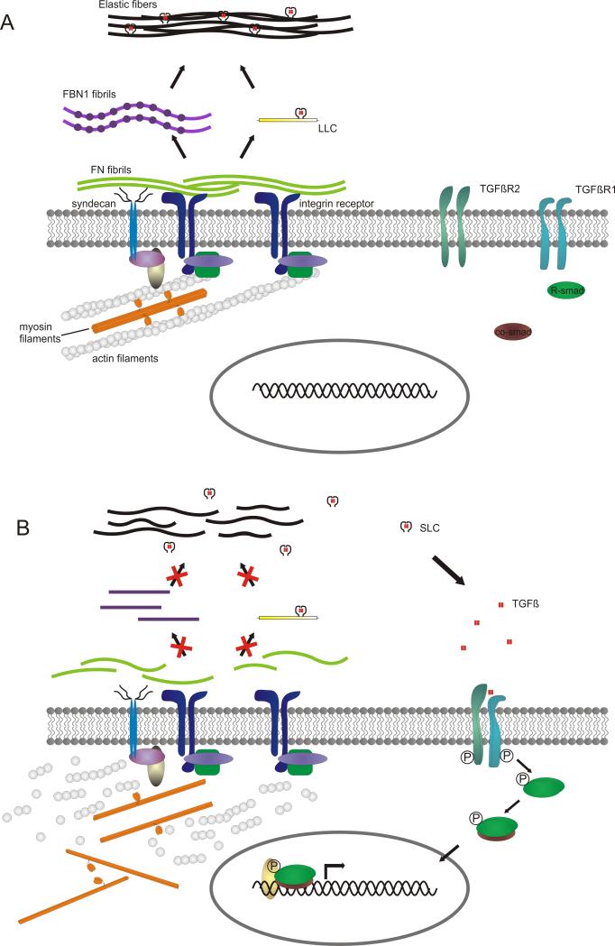
Background: Thoracic aortic aneurysm/dissection (TAAD) is a common phenotype that may occur as an isolated manifestation or within the constellation of a defined syndrome. In contrast to syndromic TAAD, the elucidation of the genetic basis of isolated TAAD has only recently started. To date, defects have been found in genes encoding extracellular matrix proteins (fibrillin-1, FBN1; collagen type III alpha 1, COL3A1), proteins involved in transforming growth factor beta (TGFβ) signaling (TGFβ receptor 1 and 2, TGFBR1/2; and SMAD3) or proteins that build up the contractile apparatus of aortic smooth muscle cells (myosin heavy chain 11, MYH11; smooth muscle actin alpha 2, ACTA2; and MYLK).

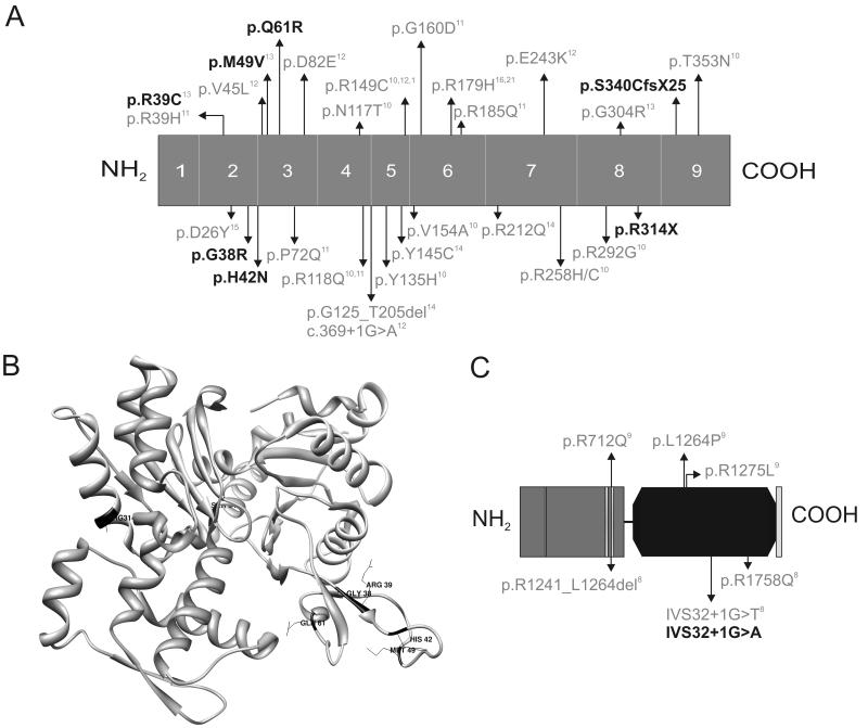
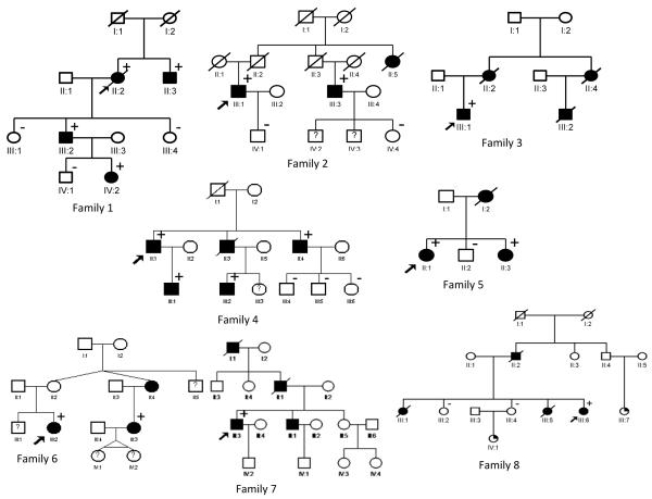
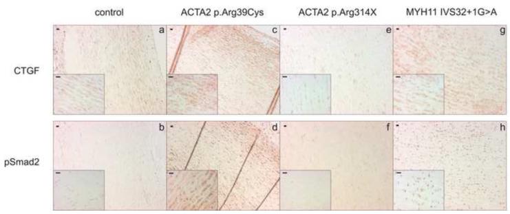
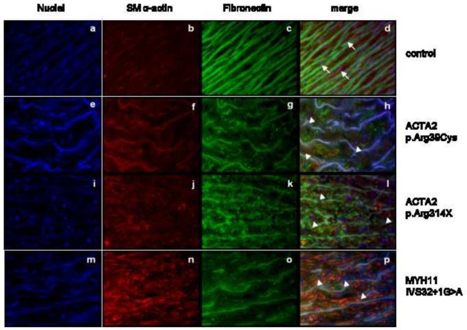
Methods and result: In 110 non-syndromic TAAD patients that previously tested negative for FBN1 or TGFBR1/2 mutations, we identified 7 ACTA2 mutations in a cohort of 43 familial TAAD patients, including 2 premature truncating mutations. Sequencing of MYH11 revealed an in frame splice-site alteration in one out of two probands with TAA(D) associated with PDA but none in the series of 22 probands from the cohort of 110 patients with non-syndromic TAAD. Interestingly, immunohistochemical staining of aortic biopsies of a patient and a family member with MYH11 and patients with ACTA2 missense mutations showed upregulation of the TGFβ signaling pathway.

Conclusions: MYH11 mutations are rare and typically identified in patients with TAAD associated with PDA. ACTA2 mutations were identified in 16% of a cohort presenting familial TAAD. Different molecular defects in TAAD may account for a different pathogenic mechanism of enhanced TGFβ signaling.


Figures

Figure 1
Figure 1
Schematic overview of the ACTA2 (A,B) and MYH11 (C) protein. A. ACTA2 mutations previously described in the literature are marked in grey and the mutations we detected here are indicated in black. The numbers 1-9 indicate the exons. B. The ACTA2 mutations identified here (black) are mapped onto the crystal structure of actin (Protein Data Bank ID 1LVT). C. MYH11 mutations previously described in literature are indicated in grey and the mutation described in this article is indicated in black. The grey amino terminal part of the schematic protein represents the myosin head-like domain with 1 ATP binding site and 2 predicted actin binding sites, the black domain is the coiled-coil domain of the myosin heavy chain 11 protein.
Figure 2
Figure 2
Pedigrees of 7 families with ACTA2 mutation (family 1-7) and 1 family with MYH11 mutation (family 8). Legend: black box = TAAD, arrow = proband, + = mutation present, − = mutation absent.
Figure 3
Figure 3
Immunohistochemical staining of aortic tissue in ACTA2 (c-f) and MYH11 (g-h) mutation-positive patients and controls (a-b). Increased cytoplasmatic staining of CTGF and nuclear phospho-Smad2 accumulation is seen in the aortic tissue of patients with ACTA2 missense mutation (c and d, respectively) and MYH11 mutation (g and h, respectively) as compared to the control (a and b, respectively). No increase in CTGF nor pSmad2 staining is seen in the aortic tissue of a patient with ACTA2 nonsense mutation (e and f, respectively).
Figure 4
Figure 4
Immunofluorescent staining of aortic tissue in ACTA2 (e-l) and MYH11 (m-p) mutation-positive patients and controls (a-d). Fragmented elastic fibers (FN1 - green) are seen in all patients (g,k,o) as compared to the control (c). Smooth muscle α-actin staining (red) is variable in ACTA2 mutation-positive patients (f,j) but seems to be increased in MYH11 mutation-positive patients (n) in comparison to the control (b). Smooth muscle cells in the control tissues (b) had an elongated shape (indicated with an arrow), while aortic tissue from patients (f,j,n) showed the presence of few round shaped cells (indicated with an arrowhead).
Figure 5
Figure 5
Hypothetical model of the pathogenetic mechanism of MYH11 and ACTA2 mutations. A. Situation in healthy individuals: fibronectin assembly is initiated with the binding of fibronectin dimers onto an integrin receptor and syndecan, which possibly acts as a co-receptor. Integrins and syndecan are also involved in assembly of the actin cytoskeleton. Fibronectin fibrillogenesis is promoted by the integrin/cytoskeleton complex by cell contractility, which induces conformational changes in fibronectin, allowing the association of multiple fibronectin molecules and formation of fibrils. Fibrillin-1 C-terminal association and formation of bead-like structures depends on these fibronectin fibrils. Fibronectin fibrils are also required for the sequestration of latent TGFβ, which is subsequently transferred onto the fibrillin-1 microfibrils, which are part of the elastic fibers. B. Mutations in MYH11 or ACTA2 will result in an incorrect assembly of the actin or myosin filaments, leading to a defect in fibronectin fibrillogenesis and resulting in an incorrect fibrillin-1 assembly into microfibrils and loss of the ability to sequestrate latent TGFβ. Latent TGFβ that is not incorporated into the matrix is more prone to activation and can bind to its receptors, initiating TGFβ signaling, characterized by phosphorylation of the receptor Smads (R-Smad), binding to the Co-Smad. This complex is then translocated to the nucleus and together with co-regulators can initiate the transcription of target genes. Used abbreviations: TGFβR1 (Transforming growth factor β receptor 1), TGFβR2 (Transforming growth factor β receptor 2), R-Smad (Receptor Smad), FN (fibronectin), FBN1 (fibrillin-1), LLC (large latent complex = LTBP (latent TGFβ binding protein) + LAP (latency associated peptide) + TGFβ), SLC (small latent complex = LAP + TGFβ), TGFβ (Transforming growth factor β).

References

    1. Lilienfeld DE, Gunderson PD, Sprafka JM, Vargas C. Epidemiology of aortic aneurysms: I. Mortality trends in the United States, 1951 to 1981. Arteriosclerosis. 1987;7:637–43. - PubMed
    1. Milewicz DM, Michael K, Fisher N, Coselli JS, Markello T, Biddinger A. Fibrillin-1 (FBN1) mutations in patients with thoracic aortic aneurysms. Circulation. 1996;94:2708–11. - PubMed
    1. Biddinger A, Rocklin M, Coselli J, Milewicz DM. Familial thoracic aortic dilatations and dissections: a case control study. J Vasc Surg. 1997;25:506–11. - PubMed
    1. Pannu H, Fadulu VT, Chang J, et al. Mutations in transforming growth factor-beta receptor type II cause familial thoracic aortic aneurysms and dissections. Circulation. 2005;112:513–20. - PubMed
    1. Guo D, Hasham S, Kuang SQ, et al. Familial thoracic aortic aneurysms and dissections: genetic heterogeneity with a major locus mapping to 5q13-14. Circulation. 2001;103:2461–8. - PubMed

Publication types

MeSH terms