. 2012 Jan;40(2):712-25.
doi: 10.1093/nar/gkr758. Epub 2011 Sep 21.

Human telomerase acts as a hTR-independent reverse transcriptase in mitochondria

Affiliations

Human telomerase acts as a hTR-independent reverse transcriptase in mitochondria

Nilesh K Sharma et al. Nucleic Acids Res. 2012 Jan.

Abstract

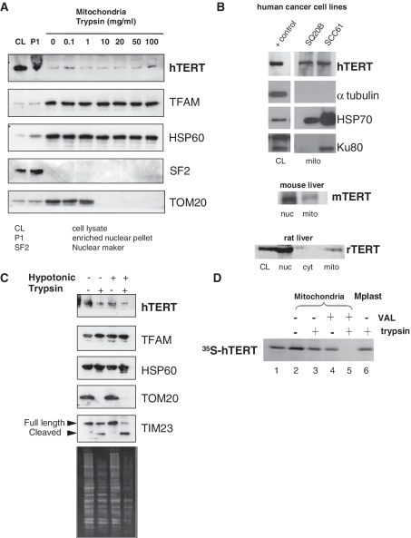
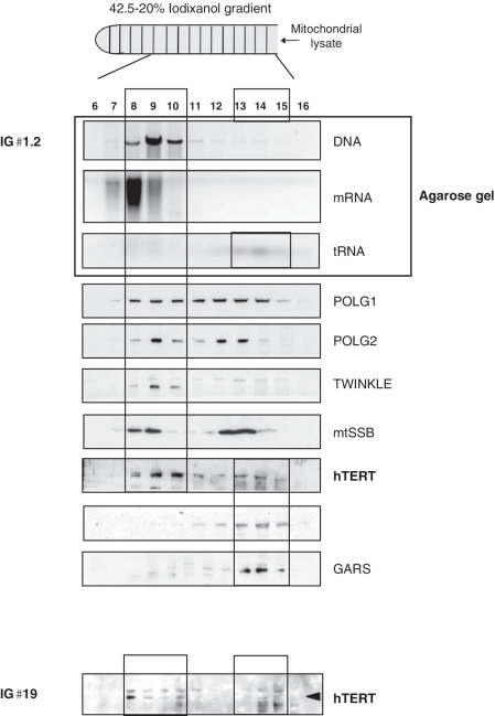
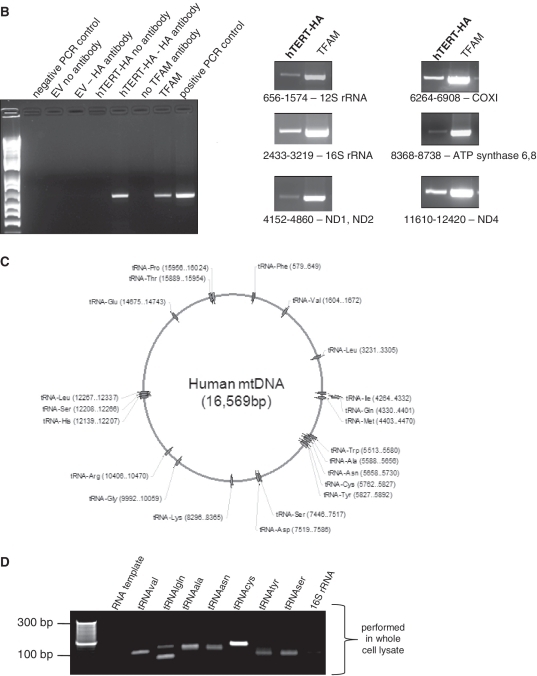
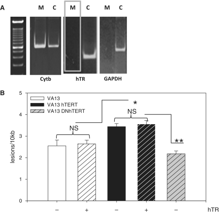
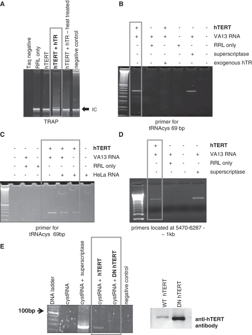
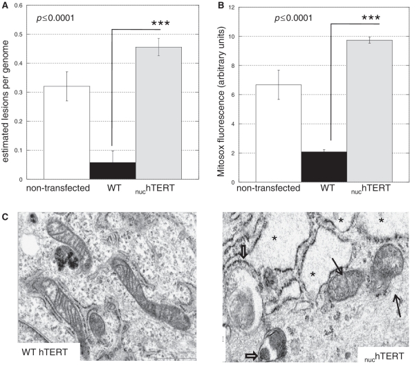
Human telomerase reverse transcriptase (hTERT) is localized to mitochondria, as well as the nucleus, but details about its biology and function in the organelle remain largely unknown. Here we show, using multiple approaches, that mammalian TERT is mitochondrial, co-purifying with mitochondrial nucleoids and tRNAs. We demonstrate the canonical nuclear RNA [human telomerase RNA (hTR)] is not present in human mitochondria and not required for the mitochondrial effects of telomerase, which nevertheless rely on reverse transcriptase (RT) activity. Using RNA immunoprecipitations from whole cell and in organello, we show that hTERT binds various mitochondrial RNAs, suggesting that RT activity in the organelle is reconstituted with mitochondrial RNAs. In support of this conclusion, TERT drives first strand cDNA synthesis in vitro in the absence of hTR. Finally, we demonstrate that absence of hTERT specifically in mitochondria with maintenance of its nuclear function negatively impacts the organelle. Our data indicate that mitochondrial hTERT works as a hTR-independent reverse transcriptase, and highlight that nuclear and mitochondrial telomerases have different cellular functions. The implications of these findings to both the mitochondrial and telomerase fields are discussed.


Figures

Figure 1.
Figure 1.
Endogenous TERT is localized to the mitochondrial matrix. (A) Mitochondria from HEK 293 cells were isolated based on differential centrifugation. The first pellet from mitochondrial isolation, consisting on nuclei and non-disrupted cells, was used as enriched nuclear pellet. Purified mitochondria were treated with increasing concentrations of trypsin to assess mitochondrial localization of proteins by western blot analysis. TFAM and HSP60 were used as mitochondrial markers while SF2 controlled for nuclear contamination. TOM20 was included to monitor trypsin efficiency. (B) Western blots showing TERT in mitochondria isolated from two human cancer cells, mouse and rat liver; CL (crude cell lysate) and nucleus: 50 µg/lane, mitochondria: 250 µg/lane. Markers for mitochondrial enrichment (HSP70) or purity (tubulin and Ku80) were included. Ku80 has a truncated version that is mitochondrial (48). Additional analysis of mitochondrial purity can be found in the Supplementary Data (Supplementary Figure S1B–S1D). (C) Mitochondria were purified from HEK 293 cells, resuspended in isotonic or hypotonic buffer and subjected to different treatments with trypsin. TOM20 was used as control for trypsin efficiency. TIM23 is present in the inner membrane with a domain facing the intermembrane space and another facing the matrix. The former is degraded by trypsin when the outer membrane is disrupted. Bottom panel shows Coomassie staining of the gel to confirm trypsin activity. (D) 35S-labeled hTERTN33-PrA was incubated with intact mitochondria in the presence of ATP (4 mM) and GTP (1 mM) at 30°C for 10 min. Valinomycin (5 mg/ml; VAL) was included as indicated. Following import, mitochondria were subjected to osmotic swelling to generate mitoplasts (lane 6). Samples were trypsin-treated and analyzed by SDS–PAGE and followed by autoradiography. Lane 1 shows 50% of 35S-labeled hTERTN33-PrA used per import assay.
Figure 2.
Figure 2.
TERT co-purifies with mitochondrial nucleic acids. (A) Purified mitochondria from HEK 293 cells were lysed and loaded onto an iodixanol gradient. IG♯1.2: no trypsin treatment was applied to mitochondria prior lysis, IG♯19: mitochondria were first treated with trypsin and then lysed. Two mitochondrial tRNA synthetases (GARS and TARS2) were used as controls for the tRNA fraction (B) mIP was performed in cells expressing hTERT-HA or empty vector (EV). Left panel shows controls as indicated in the figure; primers used encompassed region 10712–11249. Right panel shows amplification of mtDNA when using HA or TFAM for the IPs. Location of the primers and the genes present in the amplified region are shown. (C) Schematic representation of human mitochondrial genome with position of tRNA genes indicated. (D) RNA immunoprecipitations were performed in GM847 cells expressing EV or hTERT-HA. Panel shows a sample of tRNAs amplified when immunoprecipitating hTERT. First lane used RNA as template and the tRNAcys primers.
Figure 2.
Figure 2.
TERT co-purifies with mitochondrial nucleic acids. (A) Purified mitochondria from HEK 293 cells were lysed and loaded onto an iodixanol gradient. IG♯1.2: no trypsin treatment was applied to mitochondria prior lysis, IG♯19: mitochondria were first treated with trypsin and then lysed. Two mitochondrial tRNA synthetases (GARS and TARS2) were used as controls for the tRNA fraction (B) mIP was performed in cells expressing hTERT-HA or empty vector (EV). Left panel shows controls as indicated in the figure; primers used encompassed region 10712–11249. Right panel shows amplification of mtDNA when using HA or TFAM for the IPs. Location of the primers and the genes present in the amplified region are shown. (C) Schematic representation of human mitochondrial genome with position of tRNA genes indicated. (D) RNA immunoprecipitations were performed in GM847 cells expressing EV or hTERT-HA. Panel shows a sample of tRNAs amplified when immunoprecipitating hTERT. First lane used RNA as template and the tRNAcys primers.
Figure 3.
Figure 3.
hTR is not present in human mitochondria and not required for the mitochondrial effects of hTERT. (A) Whole cell lysates were treated with RNase A, washed and mitochondria were isolated. Following RNA extraction and DNase I treatment, RT-PCR was performed using 500 ng of total RNA. M = mitoplast, C = crude extract. (B) VA13 cells were infected with hTR lentivirus and/or WT hTERT or DNhTERT, submitted to treatment with 200 µM H2O2 for 60 min, and mtDNA damage analyzed by QPCR. Data are the mean of three independent experiments ± SEM. NS = not significant. No statistical difference was observed between DNhTERT and VA13 (P = 0.35).
Figure 4.
Figure 4.
hTERT drives reverse transcription independent of hTR. hTERT translated in RRL was used for first strand cDNA synthesis. (A) TRAP was performed with RRL-translated hTERT to confirm its catalytic activity. Negative control omitting Taq polymerase in the PCR was also included. (B–D) First strand cDNA synthesis reactions were performed using RNA from HeLa (hTR-positive) or VA13 (devoid of hTR) cells and random hexamers. Commercially available RT was used as control. (E) The mitochondrial tRNAcys was transcribed in vitro and the RNA used as template for first strand cDNA synthesis. WT and catalytically inactive (DN) hTERT were translated in RRL and used in the reactions. The commercially available RT was used as control. Western blots (right panel) show RRL-translated proteins; only WT hTERT is FLAG-tagged (46).
Figure 5.
Figure 5.
Lack of hTERT specifically in mitochondria negatively impacts the organelle. (A) MtDNA integrity was measured using QPCR (31–33), and estimated lesion frequency/genome calculated. Results represent the mean of six independent analyses, three in the NHF background and three in the GM7532. Since results were similar the data have been pooled. Error bars represent ± standard error of the mean (SEM). Statistical significance was evaluated using unpaired Student's t-test. (B) Mitochondria superoxide generation was evaluated using the fluorescent probe Mitosox by flow cytometry. Data represent three independent experiments. Statistical significance was evaluated using ANOVA. (C) Non-transfected parental (data not shown), WT- and nuchTERT-expressing cells were evaluated for their subcellular ultrastructures using electron microscopy. At least two independent slides were analyzed per cell type; approximately 20 cells were analyzed per slide. Data shown are representative; criteria to define damaged cellular components are described in the ‘Materials and Methods’ section. Thin black arrows indicate damaged mitochondria and thick black arrows show autophagosomes. The presence of several vacuoles in the cytoplasm is evident in the mutant cells (see asterisks).

References

    1. Santos JH, Meyer JN, Skorvaga M, Annab LA, Van Houten B. Mitochondrial hTERT exacerbates free radical-mediated mtDNA damage. Aging Cell. 2004;6:399–411. - PubMed
    1. Santos JH, Meyer JN, Van Houten B. Mitochondrial localization of hTERT as a determinant for hydrogen peroxide-induced mtDNA damage and apoptosis. Human Mol. Gen. 2006;15:1757–1768. - PubMed
    1. Ahmed S, Passos JF, Birket MJ, Beckmann T, Brings S, Peters H, Birch-Machin MA, von Zglinicki T, Saretzki G. Telomerase does not counteract telomere shortening but protects mitochondrial function under oxidative stress. J. Cell. Sci. 2008;121:1046–1053. - PubMed
    1. Haendeler J, Dröse S, Büchner N, Jakob S, Altschmied J, Goy C, Spyridopoulos I, Zeiher AM, Brandt U, Dimmeler S. Mitochondrial telomerase reverse transcriptase binds to and protects mitochondrial DNA and function from damage. Arterioscler. Thromb. Vasc. Biol. 2009;6:929–935. - PubMed
    1. Maida Y, Yasukawa M, Furuuchi M, Lassmann T, Possemato R, Okamoto N, Kasim V, Hayashizaki Y, Hahn WC, Masutomi K. An RNA-dependent RNA polymerase formed by TERT and the RMRP RNA. Nature. 2009;461:230–235. - PMC - PubMed

Publication types