Review
. 2012 Jan 1;4(1):a004903.
doi: 10.1101/cshperspect.a004903.

Overview of the matrisome--an inventory of extracellular matrix constituents and functions

Affiliations
Review

Overview of the matrisome--an inventory of extracellular matrix constituents and functions

Richard O Hynes et al. Cold Spring Harb Perspect Biol. .

Abstract

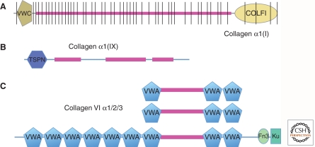
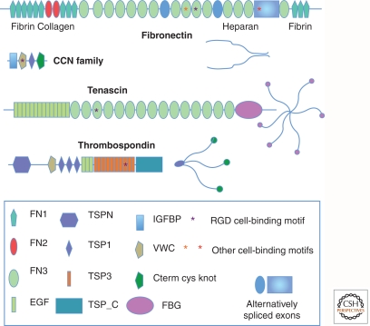
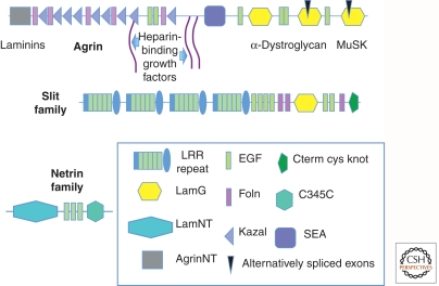
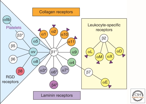
Completion of genome sequences for many organisms allows a reasonably complete definition of the complement of extracellular matrix (ECM) proteins. In mammals this "core matrisome" comprises ∼300 proteins. In addition there are large numbers of ECM-modifying enzymes, ECM-binding growth factors, and other ECM-associated proteins. These different categories of ECM and ECM-associated proteins cooperate to assemble and remodel extracellular matrices and bind to cells through ECM receptors. Together with receptors for ECM-bound growth factors, they provide multiple inputs into cells to control survival, proliferation, differentiation, shape, polarity, and motility of cells. The evolution of ECM proteins was key in the transition to multicellularity, the arrangement of cells into tissue layers, and the elaboration of novel structures during vertebrate evolution. This key role of ECM is reflected in the diversity of ECM proteins and the modular domain structures of ECM proteins both allow their multiple interactions and, during evolution, development of novel protein architectures.


Figures

Figure 1.
Figure 1.
Examples of collagen structures. (A) Collagen I is a fibrillar collagen with a continuous collagen domain of around 1000 amino acids (fuschia) comprising Gly-X-Y repeats that form a triple helix. It is encoded by multiple exons (note vertical lines) that are variants of a primordial exon encoding six such repeats. The collagen domain is flanked by amino- and carboxy-terminal noncollagenous domains that are removed by proteolysis to allow fibrillogenesis of the mature collagen. The VWC domain in this and other fibrillar collagens can be alternatively spliced and binds bone morphogenetic proteins (BMPs). (B) Collagen IX is a FACIT collagen (fibril-associated collagen with interrupted triple helix); the interruptions in the collagen domain allow bending. This and other FACIT collagens associate with fibrillar collagens and their amino-terminal domains extend out from the fibrils and presumably function as protein-binding domains. (C) Collagen VI is a heterotrimer of three related subunits, one of which is much longer and forms globular heads at each end. VWA domains are commonly protein-binding domains and probably allow interactions with other proteins during the formation of short fibrils by collagen VI.
Figure 2.
Figure 2.
Examples of characteristic ECM glycoprotein structures. Note the multidomain structure of these ECM glycoproteins. Each domain is typically encoded by a single exon or a small set of exons. This has allowed shuffling of domains into different combinations during evolution. Individual domains are specialized for binding different proteins, as indicated for fibronectin. Some domains are alternatively spliced, as noted for fibronectin, and is also true for tenascin (not shown). Cell-binding motifs such as RGD and LGV are indicated by asterisks. Fibronectin dimerizes through disulfide bonding at the carboxyl terminus, whereas thrombospondin and tenascin form trimers and hexamers, respectively, through coiled coil domains and disulfide bonds near the amino terminus. The appearances of the intact protein multimers (as would be seen by electron microscopy) are diagrammed. Note that growth factor-binding domains (IGFBP, VWC, and others) are included in many ECM proteins. CYR61 is shown as a representative member of the CCN family (see Table 2), small ECM proteins that contain integrin-binding motifs and growth-factor-binding domains (IGFBP and VWC) and are known to regulate growth factor functions (Chen and Lau 2009) as are the larger proteins shown.
Figure 3.
Figure 3.
Glycoproteins with special roles in the nervous system. These three proteins are involved in synapse formation (Agrin) and in axonal guidance (Slits and Netrins). Sites for binding other ECM proteins (laminins), growth factors, and cell-surface receptors (α-dystroglycan and MuSK) are indicated for agrin. Slit also contains known GF-binding domains (Foln). Agrin has two small alternatively spliced exons that markedly affect its functions, a characteristic of many ECM proteins. Unlike many ECM proteins, the major receptors for these three proteins are not integrins. Slit family proteins bind to Robo receptors, whereas Netrins bind to Unc5 and DCC receptors. The functions of these ECM-receptor pairs in the CNS are discussed in Barros et al. (2011) but they also function in other aspects of cell and tissue pattern regulation during development. They are evolutionarily ancient and are conserved in all bilaterial phyla.
Figure 4.
Figure 4.
Integrin receptors for ECM proteins. The diversity of integrin subunits and their interactions. Shown are the mammalian integrins, separated by color coding into subsets of closely related subunits. The RGD- binding (blue) and laminin-binding (purple) subclasses are evolutionarily very ancient and found in all metazoan phyla, but they have diverged into clades in the vertebrate lineage. The α4/α9 clade (green) is vertebrate-specific. Two subclasses of chordate α subunits have inserted I domains (purple arrowheads); they include collagen-specific integrins (orange) and a set of α subunits confined to leukocytes (yellow). Some subunits show alternatively spliced isoforms (*). The leukocyte integrins bind predominantly to cell-surface counter-receptors, whereas integrins containing either the β1 or αv subunits bind predominantly to extracellular matrix (ECM) proteins, although within each class there are exceptions to these generalizations and it is worth noting that most integrins are capable of binding multiple ligands, and there are many others beyond those shown here (Humphries et al. 2006). Many, if not all, αv integrins are also capable of activating TGF-β. Most β subunits are highly related (white) and bind to talin and related proteins (Campbell and Humphries 2011; Wickström et al. 2011), whereas the β4 subunit instead binds to intermediate filaments through specific linker proteins and the β8 subunit binds to band 4.1 proteins instead of to talin. (figure modified from Hynes 2002).
Figure 5.
Figure 5.
Evolution of ECM proteins. The figure outlines the main phylogenetic lineages (although the branch lengths are not drawn to scale), and illustrates the evolution of complexity of the matrisome and ECM during evolution. The inferred basal bilaterian had a core of ECM proteins including the basement membrane toolkit and some other ECM proteins (not all of which are shown) that have been retained in later-developing taxa, including the two main branches of metazoa (protostomes and deuterostomes). More primitive taxa had some, but not all, of these ECM proteins. During evolution of protostomes, there was modest expansion of the number of ECM genes/proteins mostly comprising taxon-specific expansions of ECM protein families by gene duplication and divergence, with some exon shuffling. A similar modest expansion occurred during evolution of the deuterostome lineage—first known acquisitions of novel ECM proteins of interest are noted in green. During evolution of the vertebrate subphylum, there was a major increase in ECM protein diversity, probably related to two whole genome duplications that occurred in that lineage. This expansion included expansion and diversification of preexisting ECM protein families, and also the development of novel protein architectures by shuffling of domains and the inclusion of novel domains (e.g., FN1, FN2, LINK). Some examples of such novel ECM proteins are indicated. As discussed in the text, this large expansion and diversification of the matrisome in vertebrates is presumably linked to novel structures such as neural crest and endothelial-lined vasculature as well as connective tissues such as cartilage, bones, and teeth, and also the development of more complex nervous and immune systems.

References

    1. Adams J, Engel J 2007. Bioinformatic analysis of adhesion proteins. In Methods in molecular biology, pp. 147–172 Humana Press, New York - PubMed
    1. Adams JC, Lawler J 2011. The thrombospondins. Cold Spring Harb Perspect Biol 10.1101/cshperspect. a009712 - DOI - PMC - PubMed
    1. Apte SS 2009. A disintegrin-like and metalloprotease (reprolysin-type) with thrombospondin type 1 motif (ADAMTS) superfamily: Functions and mechanisms. J Biol Chem 284: 31493–31497 - PMC - PubMed
    1. Aszódi A, Legate KR, Nakchbandi I, Fässler R 2006. What mouse mutants teach us about extracellular matrix function. Annu Rev Cell Dev Biol 22: 591–621 - PubMed
    1. Aumailley M, Bruckner-Tuderman L, Carter WG, Deutzmann R, Edgar D, Ekblom P, Engel J, Engvall E, Hohenester E, Jones JCR, et al. 2005. A simplified laminin nomenclature. Matrix Biol 24: 326–332 - PubMed

Publication types

LinkOut - more resources