

The clinical relevance of maintaining the functional integrity of the stratum corneum in both healthy and disease-affected skin
- PMID: 21938268
- PMCID: PMC3175800
The clinical relevance of maintaining the functional integrity of the stratum corneum in both healthy and disease-affected skin
Abstract
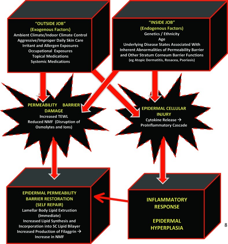
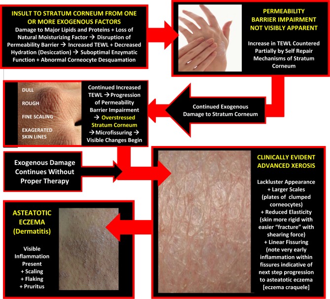
It has been recognized for approximately 50 years that the stratum corneum exhibits biological properties that contribute directly to maintaining and sustaining healthy skin. Continued basic science and clinical research coupled with keen clinical observation has led to more recent recognition and general acceptance that the stratum corneum completes many vital "barrier" tasks, including but not limited to regulating epidermal water content and the magnitude of water loss; mitigating exogenous oxidants that can damage components of skin via an innate antioxidant system; preventing or limiting cutaneous infection via multiple antimicrobial peptides; responding via innate immune mechanisms to "cutaneous invaders" of many origins, including microbes, true allergens, and other antigens; and protecting its neighboring cutaneous cells and structures that lie beneath from damaging effects of ultraviolet radiation. Additionally, specific abnormalities of the stratum corneum are associated with the clinical expression of certain disease states. This article provides a thorough "primer" for the clinician, reviewing the multiple normal homeostatic functions of the stratum corneum and the cutaneous challenges that arise when individual functions of this thin yet very active epidermal layer are compromised by exogenous and/or endogenous factors.
Figures
References
-
- Kligman AM. A brief history of how the dead stratum corneum became alive. In: Elias PM, Feingold KR, editors. Skin Barrier. New York: Taylor & Francis; 2006. pp. 15–24.
-
- Elias PM. The epidermal permeability barrier: from Saran Wrap to biosensor. In: Elias PM, Feingold KR, editors. Skin Barrier. New York: Taylor & Francis; 2006. pp. 25–32.
-
- Proksch E, Elias PM. Epidermal barrier in atopic dermatitis. In: Bieber T, Leung DYM, editors. Atopic Dermatitis. New York: Marcel Dekker; 2002. pp. 123–143.
-
- Del Rosso JQ. Understanding skin cleansers and moisturizers: the correlation of formulation science with the art of clinical use. Cosmet Dermatol. 2003;16:19–31.
-
- DiNardo A, Wertz PW. Atopic dermatitis. In: Leyden JJ, Rawlings AV, editors. Skin Moisturization. 1st ed. New York: Marcel Dekker; 2002. pp. 165–178.
LinkOut - more resources
Full Text Sources
Other Literature Sources