. 2011 Dec;31(6):1045-53.
doi: 10.1007/s10875-011-9589-4. Epub 2011 Sep 22.

Developmental, malignancy-related, and cross-species analysis of eosinophil, mast cell, and basophil siglec-8 expression

Affiliations

Developmental, malignancy-related, and cross-species analysis of eosinophil, mast cell, and basophil siglec-8 expression

Sherry A Hudson et al. J Clin Immunol. 2011 Dec.

Abstract

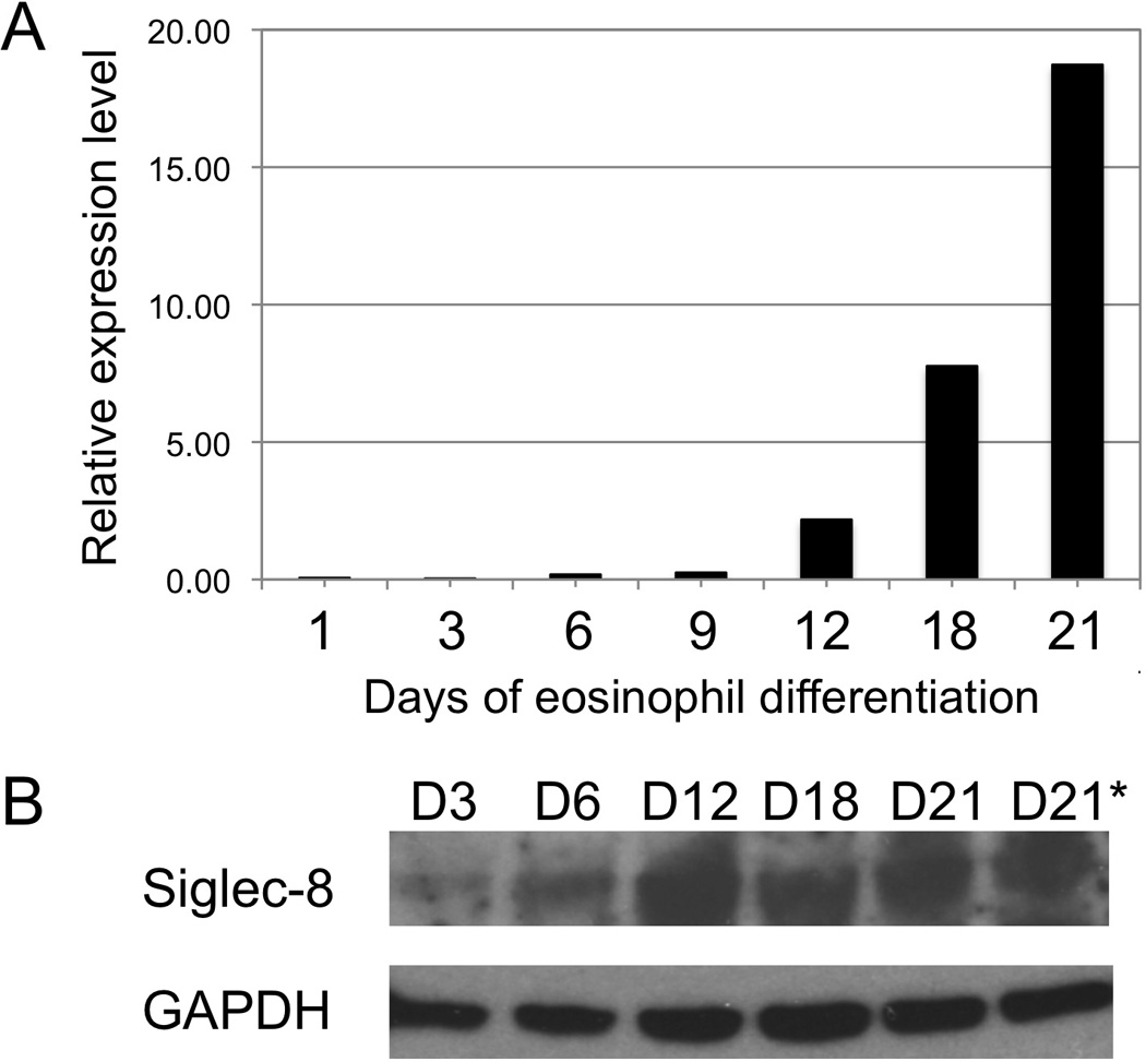
Objective: The aim of this study is to determine when during hematopoiesis Siglec-8 gets expressed, whether it is expressed on hematologic malignancies, and if there are other non-human species that express Siglec-8.

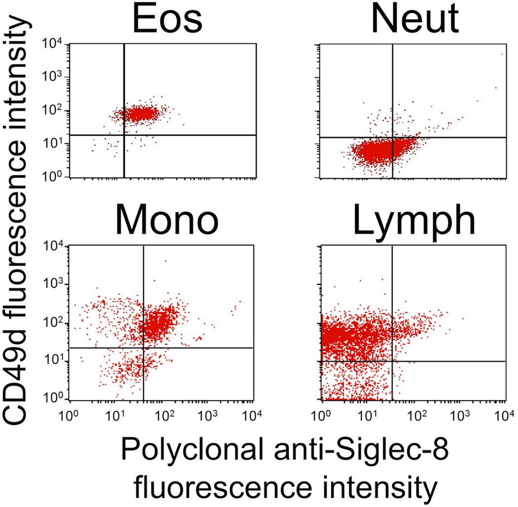
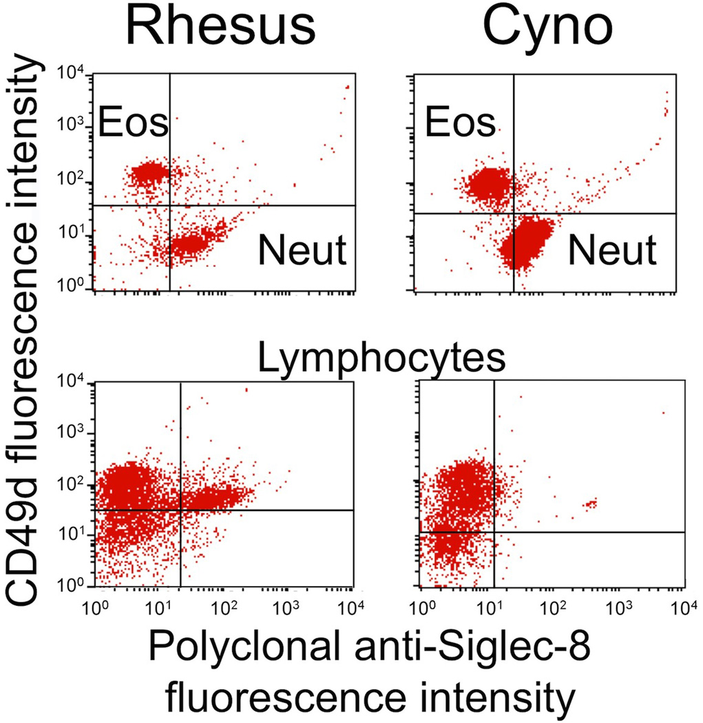
Methods: Siglec-8 mRNA and cell surface expression was monitored during in vitro maturation of human eosinophils and mast cells. Flow cytometry was performed on human blood and bone marrow samples, and on blood samples from dogs, baboons, and rhesus and cynomolgus monkeys.

Results: Siglec-8 is a late maturation marker. It is detectable on eosinophils and basophils from subjects with chronic eosinophilic leukemia, chronic myelogenous leukemia, and on malignant and non-malignant bone marrow mast cells, as well as the HMC-1.2 cell line. None of the Siglec-8 monoclonal antibodies tested recognized leukocytes from dogs, baboons, and rhesus and cynomolgus monkeys.

Conclusions: Siglec-8-based therapies should not target immature human leukocytes but should recognize mature and malignant eosinophils, mast cells, and basophils. So far, there is no suitable species for preclinical testing of Siglec-8 monoclonal antibodies.


Conflict of interest statement

The terms of this arrangement are being managed by the Johns Hopkins University in accordance with its conflict of interest policies.

Figures

Fig. 1
Fig. 1
Eosinophils express Siglec-8 late in differentiation. 2.5×10⁶ of cord blood (CB) CD34+ cells were differentiated to the eosinophil lineage in the presence of SCF, Flt3-L, IL-3, and IL-5 for 3 days, followed by IL-5 only thereafter. 5×10⁵ cells were harvested at days 3, 6, 9, 12, 15, 18, and 21. Total RNA and cell lysate were prepared at each time point. Eosinophil differentiation was confirmed using eosinophil-specific Fast Green/Neutral Red staining. a Real-time quantitative PCR analysis of Siglec-8 mRNA expression during CD34+ cell differentiation into eosinophils. b Western blot analysis of Siglec-8 protein expression during CD34+ eosinophil differentiation using 2C4 monoclonal anti-Siglec-8 antibody. GAPDH was used as a loading control (lower panel). *The duplicate Day 21 sample is from an additional CB cultured under identical conditions
Fig. 2
Fig. 2
Polyclonal Siglec-8 labeling of eosinophils (Eos), neutrophils (Neut), monocytes (Mono), and lymphocytes (Lymph) in baboon blood samples. Shown are results from a single experiment representative of three experiments. Eosinophils and neutrophils are shown separately because of the need to use different quad-stat settings due to differences in background autofluorescence seen with control antibodies. Note that the gating strategies for these experiments included light scatter to separate granulocytes from monocytes and lymphocytes, and then CD49d antibody labeling was used to distinguish eosinophils (positive) from neutrophils (negative). Also note that the CD49d+ monocytes represented contamination of the scatter gate with neutrophils
Fig. 3
Fig. 3
Polyclonal Siglec-8 labeling of eosinophils (Eos), neutrophils (Neut), monocytes (Mono), and lymphocytes (Lymph) in rhesus and cynomolgus monkey (Cyno) blood samples. Shown are results from a single experiment representative of three experiments. Note that the gating strategies for these experiments included light scatter to separate granulocytes from monocytes and lymphocytes, and then CD49d antibody labeling was used to distinguish eosinophils (positive) from neutrophils (negative)

Similar articles

Cited by

References

    1. Floyd H, Ni J, Cornish AL, Zeng Z, Liu D, Carter KC, et al. Siglec-8: a novel eosinophil-specific member of the immunoglobulin superfamily. J Biol Chem. 2000;275:861–866. - PubMed
    1. Kikly KK, Bochner BS, Freeman S, Tan KB, Gallagher KT, D'Alessio K, et al. Identification of SAF-2, a novel siglec expressed on eosinophils, mast cells and basophils. J Allergy Clin Immunol. 2000;105:1093–1100. - PubMed
    1. Bochner BS. Siglec-8 on human eosinophils and mast cells, and Siglec-F on murine eosinophils, are functionally related inhibitory receptors. Clin Exp Allergy. 2009;39:317–324. - PMC - PubMed
    1. Hudson SA, Bovin N, Schnaar RL, Crocker PR, Bochner BS. Eosinophil-selective binding and pro-apoptotic effect in vitro of a synthetic Siglec-8 ligand, polymeric 6'-sulfated sialyl Lewis X. J Pharmacol Exp Ther. 2009;330:608–612. - PMC - PubMed
    1. Nutku E, Aizawa H, Hudson SA, Bochner BS. Ligation of Siglec-8: a selective mechanism for induction of human eosinophil apoptosis. Blood. 2003;101:5014–5020. - PubMed

Publication types

MeSH terms

LinkOut - more resources