Evidence for a motor somatotopy in the cerebellar dentate nucleus--an FMRI study in humans
- PMID: 21938757
- PMCID: PMC6869902
- DOI: 10.1002/hbm.21400
Evidence for a motor somatotopy in the cerebellar dentate nucleus--an FMRI study in humans
Abstract
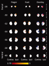
Previous anatomical studies in monkeys have shown that forelimb motor representation is located caudal to hindlimb representation within the dorso-rostral dentate nucleus. Here we investigate human dentate nucleus motor somatotopy by means of ultra-highfield (7 T) functional magnetic brain imaging (fMRI). Twenty five young healthy males participated in the study. Simple finger and foot movement tasks were performed to identify dentate nucleus motor areas. Recently developed normalization procedures for group analyses were used for the cerebellar cortex and the cerebellar dentate nucleus. Cortical activations were in good accordance with the known somatotopy of the human cerebellar cortex. Dentate nucleus activations following motor tasks were found in particular in the ipsilateral dorso-rostral nucleus. Activations were also present in other parts of the nucleus including the contralateral side, and there was some overlap between the body part representations. Within the ipsilateral dorso-rostral dentate, finger activations were located caudally compared to foot movement-related activations in fMRI group analysis. Likewise, the centre of gravity (COG) for the finger activation was more caudal than the COG of the foot activation across participants. A multivariate analysis of variance (MANOVA) on the x, y, and z coordinates of the COG indicated that this difference was significant (P = 0.043). These results indicate that in humans, the lower and upper limbs are arranged rostro-caudally in the dorsal aspect of the dentate nucleus, which is consistent with studies in non-human primates.
Copyright © 2011 Wiley Periodicals, Inc.
Figures


References
-
- Allen GI, Gilbert PF, Yin TC ( 1978): Convergence of cerebral inputs onto dentate neurons in monkey. Exp Brain Res 32: 151–170. - PubMed
-
- Asanuma C, Thach WR, Jones EG ( 1983): Anatomical evidence for segregated focal groupings of efferent cells and their terminal ramifications in the cerebellothalamic pathway of the monkey. Brain Res 286: 267–297. - PubMed
-
- Boyd LA, Winstein CJ ( 2004): Cerebellar stroke impairs temporal but not spatial accuracy during implicit motor learning. Neurorehabil Neural Repair 18: 134–143. - PubMed
-
- Cui SZ, Li EZ, Zang YF, Weng XC, Ivry R, Wang JJ ( 2000): Both sides of human cerebellum involved in preparation and execution of sequential movements. Neuroreport 11: 3849–3853. - PubMed
-
- Diedrichsen J ( 2006): A spatially unbiased atlas template of the human cerebellum. Neuroimage 33: 127–138. - PubMed
Publication types
MeSH terms
LinkOut - more resources
Full Text Sources

