. 2011 Sep 22;2(5):38.
doi: 10.1186/scrt79.

Therapeutic effects of human umbilical cord blood-derived mesenchymal stem cells after intrathecal administration by lumbar puncture in a rat model of cerebral ischemia

Affiliations

Therapeutic effects of human umbilical cord blood-derived mesenchymal stem cells after intrathecal administration by lumbar puncture in a rat model of cerebral ischemia

Jung Yeon Lim et al. Stem Cell Res Ther. .

Abstract

Introduction: Stem cell transplantation is a promising therapeutic strategy for the treatment of stroke. Mesenchymal stem cells (MSCs) are a potential cell source for clinical application because they can be easily obtained and cultivated with a high proliferative capacity. The safety and efficacy of cell therapy depends on the mode of cell administration. To determine the therapeutic potential of intrathecal administration of MSCs by lumbar puncture (LP), we administrated human umbilical cord blood-derived MSCs (hUCB-MSCs) intrathecally into the lumbar spinal cord or intravenously into the tail vein in a rat model of stroke, and then investigated whether hUCB-MSCs could enter the brain, survive, and improve post-stroke neurological functional recovery.

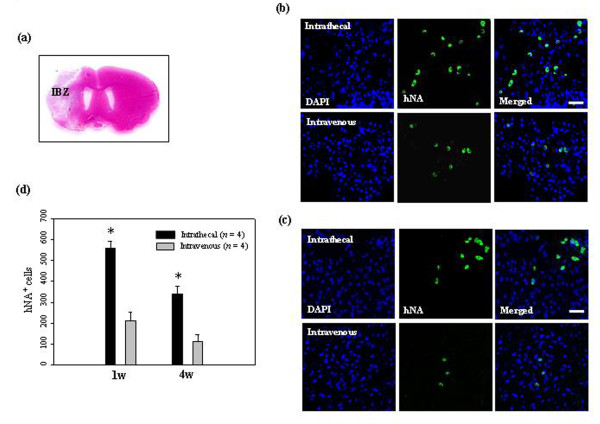
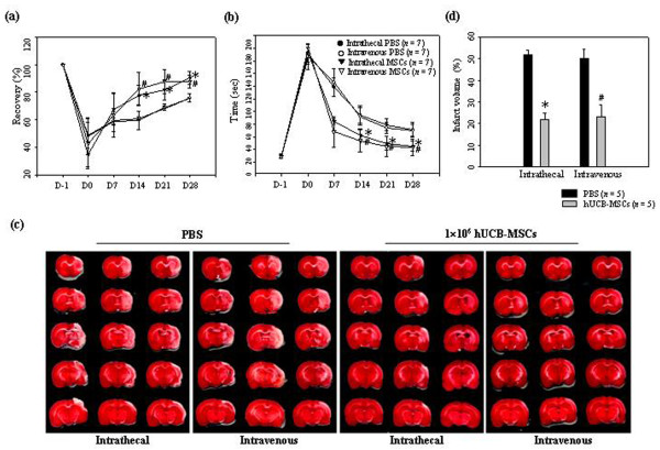
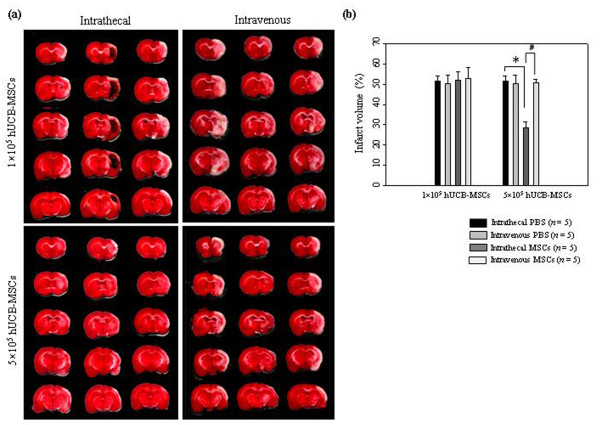
Methods: hUCB-MSCs (1.0 × 10(6)) were administrated three days after stroke induced by occlusion of the middle cerebral artery. The presence of hUCB-MSCs and their survival and differentiation in the brain tissue of the rats was examined by immunohistochemistry. Recovery of coordination of movement after administration of hUCB-MSCs was examined using a Rotarod test and adhesive-removal test on the 7th, 14th, 21st, and 28th days after ischemia. The volume of ischemic lesions seven days after the experimental procedure was evaluated using 2-3-5-triphenyltetrazolium (TTC) staining.

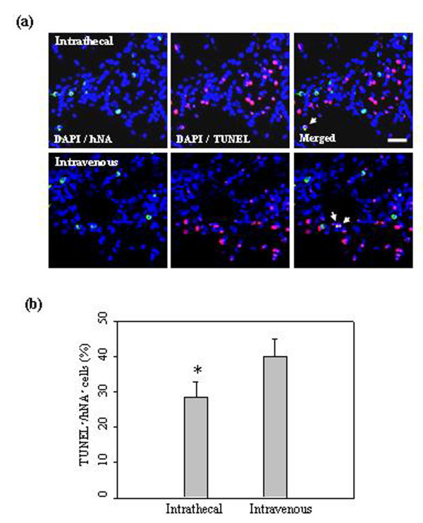
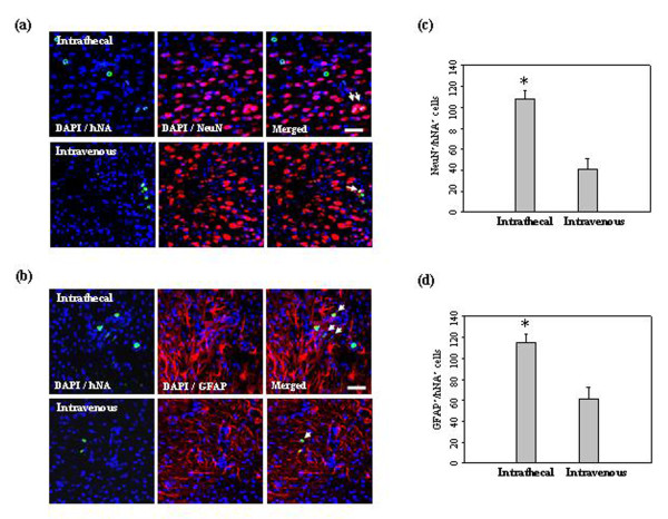
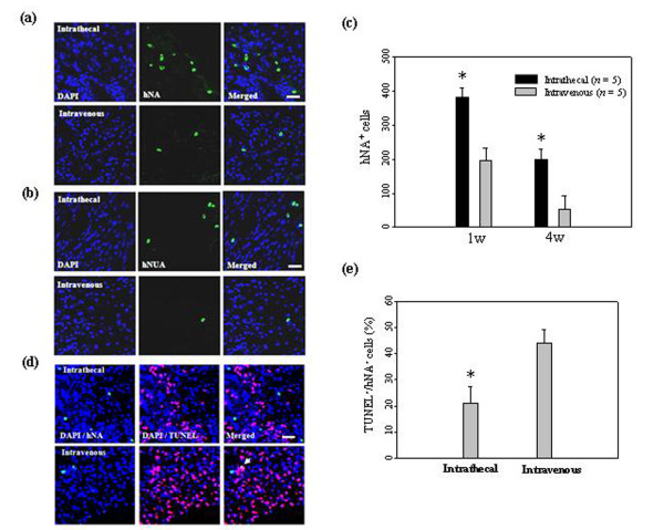
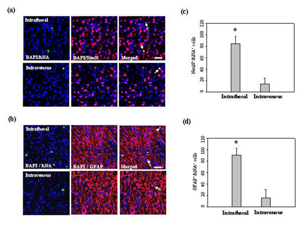
Results: Rats receiving hUCB-MSCs intrathecally by LP had a significantly higher number of migrated cells within the ischemic area when compared with animals receiving cells intravenously. In addition, many of the cells administered intrathecally survived and a subset of them expressed mature neural-lineage markers, including the mature neuron marker NeuN and glial fibrillary acidic protein, typical of astrocytes. Animals that received hUCB-MSCs had significantly improved motor function and reduced ischemic damage when compared with untreated control animals. Regardless of the administration route, the group treated with 1 × 10(6) hUCB-MSCs showed better neurological recovery, without significant differences between the two treatment groups. Importantly, intrathecal administration of 5 × 10(5) hUCB-MSCs significantly reduced ischemic damage, but not in the intravenously treated group. Furthermore, the cells administered intrathecally survived and migrated into the ischemic area more extensively, and differentiated significantly into neurons and astrocytes.

Conclusions: Together, these results indicate that intrathecal administration of MSCs by LP may be useful and feasible for MSCs treatment of brain injuries, such as stroke, or neurodegenerative disorders.


Figures

Figure 1
Figure 1
Migration of administered hUCB-MSCs into the ischemic brain. (a) Representative hematoxylin and eosin staining of coronal sections from ischemic brain. (b) At 7 days and (c) 28 days after 1 × 10⁶ hUCB-MSC administration, hUCB-MSCs were identified by the staining with human nuclei antibody (hNA, green) and the numbers of hNA-positive cells in the ischemic boundary zone (IBZ) of Ipsi hemisphere are illustrated (n = 4 per treatment group). (d) Data are presented as mean numbers of hNA-positive cells ± SD. Note that the numbers of hNA-positive cells were decreased in animals after intravenous administration compared with animals after intrathecal administration. Intrathecally treated groups showed significant differences from the intravenously treated groups in the IBZ (analysis of variance; *P < 0.05). Nuclei were counterstained with DAPI (blue). Scale bar = 20 μm.
Figure 2
Figure 2
hUCB-MSCs undergoing apoptotic cell death in the ischemic brain. (a) At seven days after 1 × 10⁶ hUCB-MSC administration, hUCB-MSCs undergoing apoptotic cell death were measured by TUNEL staining (n = 5 per treatment group). hUCB-MSCs were identified by the staining with human nuclei antibody (hNA, green). The numbers of hNA-TUNEL double-positive cells in the ipsilateral ischemic boundary zone (IBZ) are illustrated. (b) Quantitative analysis of hNA-TUNEL double-positive cells in the ipsilateral IBZ. Data from five animals are presented as mean values ± SD. There were significantly more hNA-TUNEL double-positive cells in animals after intravenous administration (analysis of variance; *P < 0.05). Nuclei were counterstained with DAPI (blue). Scale bar = 20 μm.
Figure 3
Figure 3
In vivo differentiation of hUCB-MSCs in the ischemic brain. Confocal images of the cells at four weeks after 1 × 10⁶ hUCB-MSC administration in the ischemic animal models. hUCB-MSCs were identified by the staining with human nuclei antibody (hNA, green). hUCB-MSCs survived for at least four weeks and a subset of the grafted cells expressed (a) NeuN and (b) GFAP in the ipsilateral ischemic boundary zone (IBZ). These markers were immunolabeled with red fluorescence. Quantitative analysis of (c) hNA-NeuN and (d) hNA-GFAP double-positive cells in the ipsilateral IBZ. Data are presented as mean values ± SD. Nuclei were counterstained with DAPI (blue). Scale bar, 20 μm.
Figure 4
Figure 4
Therapeutic effects of hUCB-MSC administration on recovery in the ischemic animal model. Performance in the (a) Rotarod and (b) adhesive removal tests from 1 to 28 days after ischemia. The data were collected from seven animals per group and are presented as mean values ± SD. (c) Brain slices were stained with TTC at seven days after PBS or 1 × 10⁶ hUCB-MSC administration to visualize lesions. (d) The data were collected from five animals per group and are presented as mean relative infarct volume ± SD. Statistically significant differences between the groups were determined by analysis of variance (*P < 0.05 compared with the PBS injected group intrathecally; #P < 0.05 compared with the PBS injected group intravenously).
Figure 5
Figure 5
Therapeutic effects of low-dose hUCB-MSC administration on infarction volume in the ischemic animal model. (a) Brain slices were stained with TTC at seven days after 5 × 10⁵ hUCB-MSC administration. The images show the lesion volume in hUCB-MSCs treated groups. (b) The data were collected from five animals per group and are presented as mean relative infarct volume ± SD. Statistically significant differences between the groups were determined by analysis of variance (*P < 0.05 compared with the PBS injected group intrathecally; #P < 0.05 compared with the hUCB-MSCs injected group intravenously).
Figure 6
Figure 6
Migration and survival of low-dose hUCB-MSCs in the ischemic brain. (a) At 7 days and (b) 28 days after 5 × 10⁵ hUCB-MSC administration, hUCB-MSCs were identified by the staining with human nuclei antibody (hNA, green) and the numbers of hNA-positive cells in the ischemic boundary zone (IBZ) of Ipsi hemisphere are illustrated (n = 5 per treatment group). (c) Data are presented as mean numbers of hNA-positive cells ± SD. Note that the numbers of hNA-positive cells were decreased in animals after intravenous administration compared with animals after intrathecal administration. Intrathecally treated groups showed significant differences from the intravenously treated groups in the IBZ. (d) At seven days after 5 × 10⁵ hUCB-MSC administration, hUCB-MSCs undergoing apoptotic cell death were measured by TUNEL staining. The numbers of hNA-TUNEL double-positive cells in the ipsilateral IBZ are illustrated. (e) Quantitative analysis of hNA-TUNEL double-positive cells in the ipsilateral IBZ. Data from five animals are presented as mean values ± SD. There were significantly more hNA-TUNEL double-positive cells in animals after intravenous administration (analysis of variance; *P < 0.05). Nuclei were counterstained with DAPI (blue). Scale bar = 20 μm.
Figure 7
Figure 7
In vivo differentiation of low-dose hUCB-MSCs in the ischemic brain. Confocal images of the cells at four weeks after 5 × 10⁵ hUCB-MSC administration in the ischemic animal models. hUCB-MSCs were identified by the staining with human nuclei antibody (hNA, green). A subset of the grafted cells expressed (a) NeuN and (b) GFAP in the ipsilateral ischemic boundary zone. These markers were immunolabeled with red fluorescence. Quantitative analysis of (c) hNA-NeuN and (d) hNA-GFAP double-positive cells in the ipsilateral IBZ. Data are presented as mean values ± SD. There were significantly more double-positive cells in animals after intrathecal administration (analysis of variance; *P < 0.05). Nuclei were counterstained with DAPI (blue). Scale bar = 20 μm.

References

    1. Chen J, Chopp M. Neurorestorative treatment of stroke: cell and pharmacological approaches. NeuroRx. 2006;3:466–473. doi: 10.1016/j.nurx.2006.07.007. - DOI - PMC - PubMed
    1. Caplan AI. Mesenchymal stem cells. J Orthop Res. 1991;9:641–650. doi: 10.1002/jor.1100090504. - DOI - PubMed
    1. Goodwin HS, Bicknese AR, Chien SN, Boqucki BD, Quinn CO, Wall DA. Multilineage differentiation activity by cells isolated from umbilical cord blood: expression of bone, fat, and neural markers. Biol Blood Marrow Transplant. 2001;7:581–588. doi: 10.1053/bbmt.2001.v7.pm11760145. - DOI - PubMed
    1. Pittenger MF, Mackay AM, Beck SC, Jaiswal PK, Douglas R, Mosca JD, Moorman MA, Simonetti DW, Craig S, Marshak DR. Multilineage potential of adult human mesenchymal stem cells. Science. 1999;284:143–147. doi: 10.1126/science.284.5411.143. - DOI - PubMed
    1. Deng W, Obrocka M, Fischer I, Prockop DJ. In vitro differentiation of human marrow stromal cells into early progenitors of neural cells by conditions that increase intracellular cyclic AMP. Biochem Biophys Res Commun. 2001;282:148–152. doi: 10.1006/bbrc.2001.4570. - DOI - PubMed

Publication types

LinkOut - more resources