. 2012 Jan 16;59(2):1912-23.
doi: 10.1016/j.neuroimage.2011.08.102. Epub 2011 Sep 13.

Impact of state anxiety on the interaction between threat monitoring and cognition

Affiliations

Impact of state anxiety on the interaction between threat monitoring and cognition

Jong Moon Choi et al. Neuroimage. .

Abstract

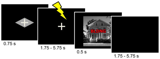
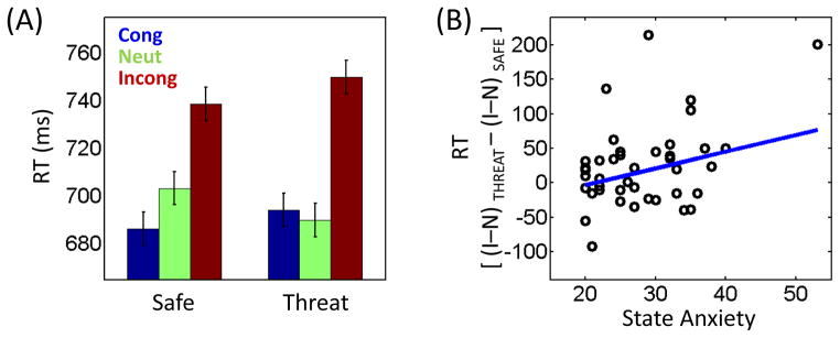
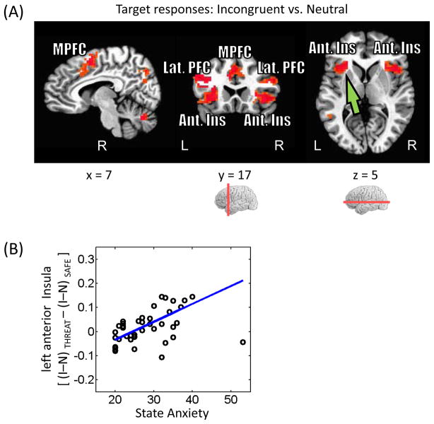
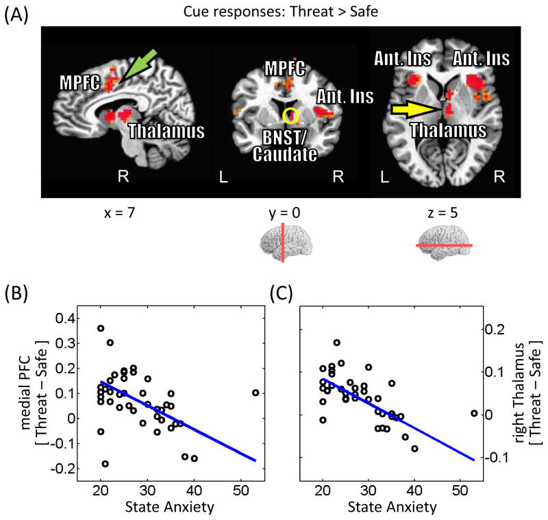
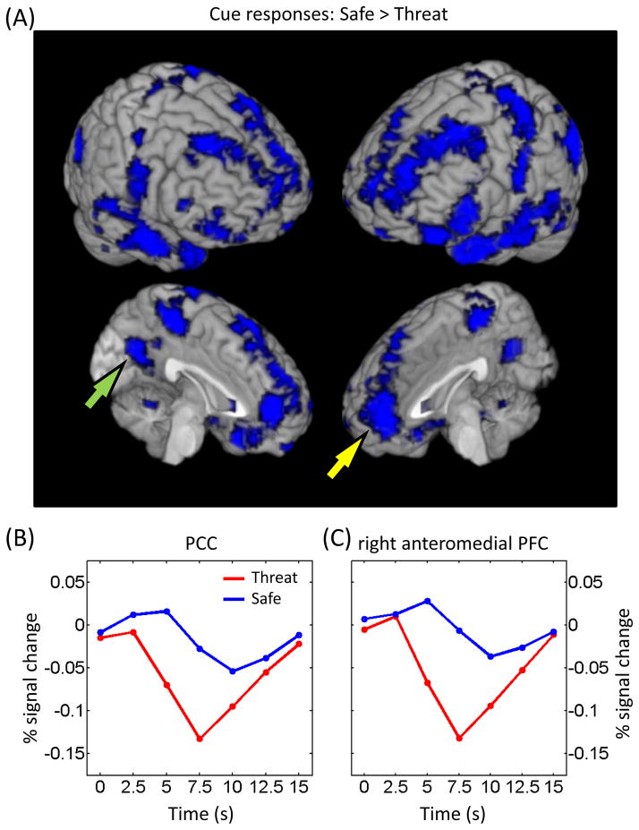
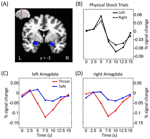
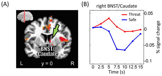
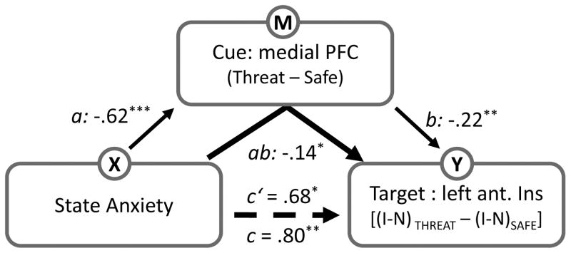
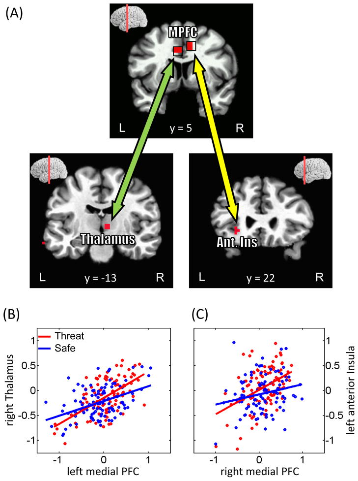
How does threat processing impact cognitive performance? To investigate this question, in the present functional magnetic resonance imaging study, participants performed a response-conflict task (neutral, congruent, and incongruent trials) that followed a variable-length shock anticipation period or a corresponding delay during which they would not be shocked. The delay period was cued by a geometric-shaped stimulus indicating whether the subject was in the safe (no shock) or threat (potential shock) condition. Behaviorally, participants showed increased reaction time interference (incongruent-neutral) during threat trials, an effect that increased as a function of state anxiety level across participants. Brain imaging data were analyzed for the cue and the subsequent target phase of the task. At the target phase, the left anterior insula exhibited interaction-type responses (i.e., increased interference during threat trials) that were positively associated with state anxiety level - a relationship that paralleled the behavioral pattern. At the cue phase, greater responses to threat vs. safe were observed in a circuit of regions, including the medial PFC, anterior insula, thalamus, and bed nucleus of the stria terminalis/caudate, which we interpreted as engaged by shock monitoring/anticipation processes. In contrast, intriguingly, greater responses to safe vs. threat at the cue phase were observed in a broader set of regions that overlapped with the "resting-state" network. Finally, a standard statistical mediation analysis revealed that the relationship between state anxiety scores and interference-related responses in the left anterior insula during the target phase was partially mediated via cue responses in the medial PFC, consistent with the idea that more anxious individuals had difficulty in engaging the medial PFC during the threat condition. Taken together, our findings suggest that threat monitoring impairs the upcoming resolution of interference. Furthermore, a confluence of effects of cognitive task condition, threat, and individual differences in state anxiety was observed in the anterior insula, a structure that is suggested to be particularly important for the interaction between emotion and cognition.


Figures

Figure 1
Figure 1
Task design. Subjects performed a response-conflict task under two contexts, safe and threat. During the threat condition (shown here), a cue stimulus (diamond) signaled that a mild electric shock could occur during the delay period following cue offset and prior to the target display. Participants were instructed about the meaning of the cue stimuli prior to task execution. During the target phase, participants were asked to indicate whether the picture contained a house or a building, while ignoring the superimposed word. During the safe condition (not shown), the trial structure was identical, except for the shape of the cue stimulus (rectangle) and the fact that shocks were never administered.
Figure 2
Figure 2
Behavioral results. (A) Participants exhibited slower responses during incongruent trials. Importantly, the amount of interference (incongruent vs. neutral) was greater when the target display was preceded by a threat relative to safe cue. Cong, congruent trials; Neut, neutral trials; Incong, incongruent trials. (B) Across participants, interaction-type scores were linearly related to state anxiety levels. The blue line indicates the robust linear regression fit. I, incongruent trials; N, neutral trials. Error bars in panel A denote the standard within-subject error term (Loftus and Masson, 1994).
Figure 3
Figure 3
Target-related responses. (A) Voxels that showed stronger responses during incongruent than neutral trials (displayed at p < 0.05, cluster-level corrected). MPFC, medial prefrontal cortex; Lat. PFC, lateral prefrontal cortex; Ant. Ins, anterior insula. (B) Across participants, interaction-type scores in the left anterior insula (see green arrow in panel A) were positively related to state anxiety levels. The blue line indicates the robust linear regression fit. I, incongruent trials; N, neutral trials.
Figure 4
Figure 4
Cue-related responses. (A) Voxels that showed stronger responses during threat than safe trials (displayed at p < 0.05, cluster-level corrected). MPFC, medial prefrontal cortex; BNST, bed nucleus of the stria terminalus (yellow circle); Ant. Ins, anterior insula. (B) Across participants, differential responses in the medial prefrontal cortex (see green arrow in panel A) were inversely related to state anxiety levels. The blue line indicates the robust linear regression fit. (C) As in (B) for the right thalamus (see yellow arrow in panel A).
Figure 5
Figure 5
Cue-related responses. (A) Voxels that showed stronger responses during safe than threat trials (displayed at p < 0.05, cluster-level corrected). The overall pattern was very similar to the one reported for the task-negative network. (B, C) Mean estimated responses from the PCC (B; see green arrow in panel A) and right anteromedial PFC (C; see yellow arrow in panel A) during safe and threat conditions. PCC, posterior cingulate cortex; PFC, prefrontal cortex.
Figure 6
Figure 6
Cue-related responses in the amygdala. (A) Voxels within the anatomically defined amygdala (as defined via the AFNI anatomical template; black outline) exhibiting stronger responses during safe vs. threat. (B) Mean estimated responses from the left and right amygdala during trials in which electrical stimulation was administered. (C, D) Mean estimated responses from the left (C) and right (D) amygdala during safe and threat conditions (as in all other analyses reported in the paper [except those of panel B], trials containing shock were discarded).
Figure 7
Figure 7
Cue-related responses in the bed nucleus of the stria terminalus (BNST)/caudate. (A) Voxels that showed stronger responses during threat than safe trials (displayed at p < 0.05, cluster-level corrected). (B) Mean estimated responses from the right BNST/caudate (see green arrows in panel A) during safe and threat conditions. At an exploratory threshold of .001 (uncorrected), we observed similar responses in the left BNST/caudate.
Figure 8
Figure 8
Mediation analysis. We tested whether the relationship between state anxiety and target responses was mediated via responses at the cue phase. For the target phase, interaction-type scores were considered, whereas differential responses were considered during the cue phase. The letters a, b, c and c’ refer to estimated path coefficients. The dotted line indicates the path coefficient was reduced after the mediator was taken into account. Ant. Ins; anterior insula. I, incongruent trials; N, neutral trials. *p < .05, **p < .01, ***p < .005.
Figure 9
Figure 9
Functional connectivity on a trial-by-trial basis. (A) Regions exhibiting stronger functional connectivity with the medial PFC during the cue phase. The top panel illustrates the ROIs employed as “seed” regions. MPFC, medial prefrontal cortex; Ant. Ins, anterior insula. (B, C) The scatter plots show trial-by-trial responses in the left medial PFC and right thalamus (B) and in the right medial PFC and left anterior insula (C). Responses to threat trials are shown in red and to safe trials are shown in blue. Robust linear fits to the data are presented in the corresponding colors. Data are illustrated for representative individuals.

References

    1. Anticevic A, Repovs G, Barch DM. Resisting emotional interference: Brain regions facilitating working memory performance during negative distraction. Cognitive, Affective, & Behavioral Neuroscience. 2010;10:159–173. - PMC - PubMed
    1. Bach DR, Flandin G, Friston KJ, Dolan RJ. Time-series analysis for rapid event-related skin conductance responses. Journal of Neuroscience Methods. 2009;184:224–234. - PMC - PubMed
    1. Banks SJ, Eddy KT, Angstadt M, Nathan PJ, Phan KL. Amygdala–frontal connectivity during emotion regulation. Social Cognitive and Affective Neuroscience. 2007;2:303–312. - PMC - PubMed
    1. Baron RM, Kenny DA. The moderator–mediator variable distinction in social psychological research: Conceptual, strategic, and statistical considerations. Journal of personality and social psychology. 1986;51:1173–1182. - PubMed
    1. Besner D, Stolz JA, Boutilier C. The stroop effect and the myth of automaticity. Psychon Bull Rev. 1997;4:221–225. - PubMed