

Default mode network connectivity predicts sustained attention deficits after traumatic brain injury
- PMID: 21940437
- PMCID: PMC6623308
- DOI: 10.1523/JNEUROSCI.1163-11.2011
Default mode network connectivity predicts sustained attention deficits after traumatic brain injury
Abstract
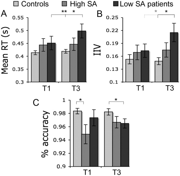
Traumatic brain injury (TBI) frequently produces impairments of attention in humans. These can result in a failure to maintain consistent goal-directed behavior. A predominantly right-lateralized frontoparietal network is often engaged during attentionally demanding tasks. However, lapses of attention have also been associated with increases in activation within the default mode network (DMN). Here, we study TBI patients with sustained attention impairment, defined on the basis of the consistency of their behavioral performance over time. We show that sustained attention impairments in patients are associated with an increase in DMN activation, particularly within the precuneus and posterior cingulate cortex. Furthermore, the interaction of the precuneus with the rest of the DMN at the start of the task, i.e., its functional connectivity, predicts which patients go on to show impairments of attention. Importantly, this predictive information is present before any behavioral evidence of sustained attention impairment, and the relationship is also found in a subgroup of patients without focal brain damage. TBI often results in diffuse axonal injury, which produces cognitive impairment by disconnecting nodes in distributed brain networks. Using diffusion tensor imaging, we demonstrate that structural disconnection within the DMN also correlates with the level of sustained attention. These results show that abnormalities in DMN function are a sensitive marker of impairments of attention and suggest that changes in connectivity within the DMN are central to the development of attentional impairment after TBI.
Figures


Comment in
-
Learning from default mode network: the predictive value of resting state in traumatic brain injury.J Neurosci. 2012 Feb 8;32(6):1915-7. doi: 10.1523/JNEUROSCI.5637-11.2012. J Neurosci. 2012. PMID: 22323703 Free PMC article. No abstract available.
References
-
- Ashburner J, Friston KJ. Voxel-based morphometry—the methods. Neuroimage. 2000;11:805–821. - PubMed
-
- Azouvi P, Couillet J, Leclercq M, Martin Y, Asloun S, Rousseaux M. Divided attention and mental effort after severe traumatic brain injury. Neuropsychologia. 2004;42:1260–1268. - PubMed
-
- Baddeley AD, Emslie H, Nimmo-Smith I. Doors and people test: a test of visual and verbal recall and recognition. Bury-St-Edmunds, UK: Thames Valley Test Company; 1994.
-
- Beckmann CF, Smith SM. Probabilistic independent component analysis for functional magnetic resonance imaging. IEEE Trans Med Imaging. 2004;23:137–152. - PubMed
-
- Beckmann CF, Jenkinson M, Smith SM. General multilevel linear modeling for group analysis in FMRI. Neuroimage. 2003;20:1052–1063. - PubMed
Publication types
MeSH terms
Grants and funding
LinkOut - more resources
Full Text Sources
Medical