. 2011 Oct 4;108(40):16759-64.
doi: 10.1073/pnas.1110904108. Epub 2011 Sep 21.

A nucleolar protein, H19 opposite tumor suppressor (HOTS), is a tumor growth inhibitor encoded by a human imprinted H19 antisense transcript

Affiliations

A nucleolar protein, H19 opposite tumor suppressor (HOTS), is a tumor growth inhibitor encoded by a human imprinted H19 antisense transcript

Patrick Onyango et al. Proc Natl Acad Sci U S A. .

Abstract

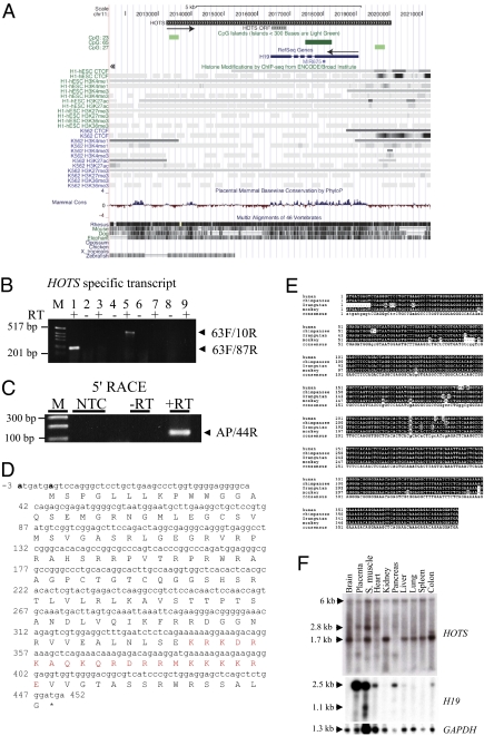
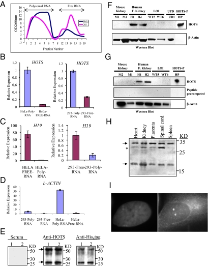
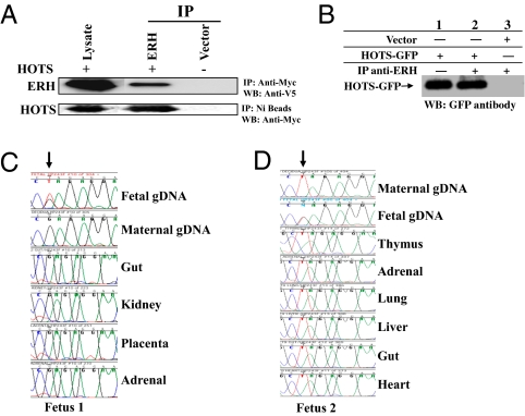
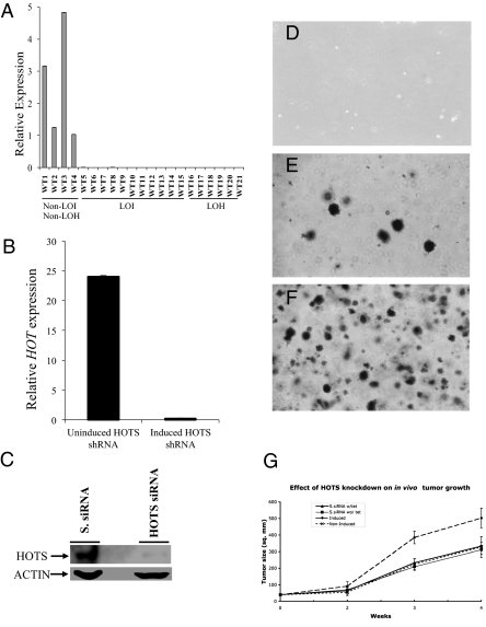
The H19 gene, which localizes within a chromosomal region on human chromosome 11p15 that is commonly lost in Wilms tumor (WT), encodes an imprinted untranslated RNA. However, the biological significance of the H19 noncoding transcript remains unresolved because replacement of the RNA transcript with a neocassette has no obvious phenotypic effect. Here we show that the human H19 locus also encodes a maternally expressed, translated gene, antisense to the known H19 transcript, which is conserved in primates. This gene, termed HOTS for H19 opposite tumor suppressor, encodes a protein that localizes to the nucleus and nucleolus and that interacts with the human enhancer of rudimentary homolog (ERH) protein. WTs that show loss of heterozygosity of 11p15 or loss of imprinting of IGF2 also silence HOTS (7/7 and 10/10, respectively). Overexpression of HOTS inhibits Wilms, rhabdoid, rhabdomyosarcoma, and choriocarcinoma tumor cell growth, and silencing HOTS by RNAi increases in vitro colony formation and in vivo tumor growth. These results demonstrate that the human H19 locus harbors an imprinted gene encoding a tumor suppressor protein within the long-sought WT2 locus.


Conflict of interest statement

The authors declare no conflict of interest.

Figures

Fig. 1.
Fig. 1.
HOTS is a primate-conserved, ubiquitous transcript antisense to H19. (A) Genomic organization of HOTS and H19 genes on human chromosome 11p15.5. The direction of HOTS and H19 transcriptional orientation is shown by black arrows. HOTS ORF is shown. The display is populated with CpG island (green) CTCF-binding sites, ENCODE histone modification marks, and mammalian DNA sequence conservation for the region [University of California at Santa Cruz (UCSC) on Human Genome Sequence: Feb. 2009 (GRCh37/hg19) Assembly, http://genome.ucsc.edu/]. (B) RT-PCR amplification of HOTS using strand-specific RT primer 1119R located within the first intron of H19, followed by amplification with intronic PCR primer pairs 63F/87R (lanes 1 and 2) and 63F/10R (lanes 5 and 6). RT-PCR amplification of H19 using strand-specific RT primer 1F, followed by intronic PCR primer pair 63F/87R (lanes 3 and 4) or 63F/10R (lanes 7 and 8). (Lane 9) No RT primer included but RT enzyme and 63F/87R primer pair. (C) 5′ RACE using primer 4534R for RT and primers 44R and PCR anchor primer. M, 1-kb DNA ladder; NTC, no template control. (D) Sequence for the HOTS ORF, nuclear localization signal shown in red type and the putative Kozak consensus sequences at −3 (A) and +4 (A) in boldface type. (E) Multiple sequence alignment of predicted HOTS primates sequences from human, chimpanzee, orangutan, and monkey. Sequence areas depicted against a black background represent identity, and small letters in the consensus sequence at the bottom show areas of sequence variability. (F) HOTS and H19 human multiple tissues Northern blots. (Top) HOTS. (Middle) H19. (Bottom) GAPDH.
Fig. 2.
Fig. 2.
HOTS is a polysome-associated RNA encoding a nucleolar protein. (A) Polysomal and free total cellular RNA fractionated by sucrose gradient centrifugation from homogenates of HeLa (blue) and HEK293 (pink) human cells. (B) Strand-specific quantitative real-time PCR amplification of HOTS transcripts shows enrichment in polysomes from HeLa and HEK293 cells, in contrast to (C) H19, which shows enrichment in the free RNA fraction, with (D) β-actin a positive control for polysome enrichment. (B–D) Analyses were performed in triplicate (n = 6). (E) Western blots of purified His6-tag HOTS protein loaded in duplicate lanes using preimmune serum (Left), HOTS antibody (Center), and His6-tag antibody (Right). Both the HOTS and His6-tag antibodies detect the expected recombinant protein (arrows) of 26 kDa (17 kDa of predicted HOTS polypeptide sequence and 9 kDa of His6 plus trailing vector amino acid sequences) and a dimer of 52 kDa. (F) Western blot with anti-HOTS antibody on protein extracts from mouse kidney (M1 and M2), human fetal kidney (H1 and H2), WTs with LOI (WT5 and WT6), a BWS sample with chromosome 11p UPD (UD1), and purified HOTS protein (HP). The LOI and UD samples are negative controls with loss of expression of the maternally expressed HOTS. β-Actin antibody was used on the same blot as a loading control. (G) Western blot similar to the previous but precompeted with HOTS purified protein (1 μg/mL). (H) Western blots of a nondenaturing gel using anti-HOTS antibody on human fetal tissues. Arrows indicate the HOTS monomer of 17 kDa and a dimer of 34 kDa; asterisk indicates a 29-kDa band that might represent an isoform or posttranslationally modified protein abundant in the spinal cord. (I) Subcellular localization of native HOTS protein to the nucleolus using anti-HOTS antibody on human SiHa cells. (Left) DAPI stained nucleus. (Right) HOTS antibody image superimposed on the DAPI stain.
Fig. 3.
Fig. 3.
HOTS interacts with ERH in vivo and is imprinted. (A) (Upper) Immunoprecipitation (IP) with anti–myc-tag antibodies specific to myc-tagged HOTS from HEK293 cells cotransfected with myc-tagged HOTS and His6/V5-tagged ERH. V5 antibody was used to detect ERH on the Western blot (WB). (Lower) IP with nickel beads specific for His-tagged ERH from HEK293 cells transfected with myc-tagged HOTS and His6/V5-tagged ERH; myc-tagged antibody was used to detect HOTS by WB. (B) Immunoprecipitation of HOTS from protein extracts of HEK293 cells transfected with HOTS tagged with GFP and immunoprecipitated using ERH antibody. Ten percent (30 μg) of HEK293 input extract was loaded in lane 1. Anti-GFP antibody was used for Western blot. (C) Strand-specific cDNA synthesis was carried out to study HOTS expression in the mother and in fetal tissues of fetus 1. The polymorphism is shown by an arrow. The maternal decidua and fetal genomic DNA sequence is included to distinguish the origin of the expressed allele. Note the exclusive expression the maternal G allele in all fetal tissues. (D) Maternal T allele expression in all fetus 2 tissues (indicated by an arrow). Note that the fetus genomic DNA is heterozygous and therefore informative for allele-specific gene expression. All cDNA synthesis was performed with gene-specific primers that produced only HOTS transcripts.
Fig. 4.
Fig. 4.
HOTS is lost in LOH and LOI WT and suppresses tumor cell growth in vitro and in vivo. (A) Quantitative real-time expression analysis of HOTS in WT samples shows loss of expression in LOI (WT5–14) and LOH (WT15–21) cases, but not in non-LOH, non-LOI cases (WT1–4). Error bars (SD) are plotted but are too small to see. (B) Tetracycline-inducible knockdown of HOTS by HOTS shRNA in HeLa cells. Induction with tetracycline activates the HOTS shRNA-expressing plasmid, leading to silencing of HOTS. (C) Western blot showing siRNA knockdown of HOTS with HOTS siRNA but not with scrambled siRNA (S. siRNA). β-Actin was used as a loading control. (D–F) HeLa tumor cell growth assayed on soft agar, showing that growth is inhibited by HOTS expressed from a tetracycline-inducible vector after Zeocin selection. (D) Nontransfected HeLa cells, selected with 0.5 mg/mL Zeocin. No visible colonies were observed due to cell death from Zeocin drug selection. (E) HeLa cell colonies from a culture that was not induced to express anti-HOTS RNAi. (F) Fifteen-fold more HeLa cell colonies occur when cells are induced by tetracycline to express anti-HOTS RNAi. (Scale bar, D–F: 0.3 mm.) n = 3. (G) Increased tumor cell growth in nude mice upon RNAi knockdown of HOTS. n = 6. Mean tumor volume was plotted against time. Statistically significant difference in tumor area between the HOTS knockdown animals (Induced, •) and the HOTS-expressing animals (Non-Induced, X-dotted line) was scored (P < 0.01 at 3 wk and P < 0.02 at 4 wk, Student's t-test). As controls, we included scrambled siRNA (S.siRNA) tetracycline-induced (▲, w/tet) and Non-Induced S.siRNA (■, wo/tet) HeLa transfected cells.

Comment in

References

    1. Arney KL. H19 and Igf2: Enhancing the confusion? Trends Genet. 2003;19:17–23. - PubMed
    1. Tremblay KD, Duran KL, Bartolomei MS. A 5′ 2-kilobase-pair region of the imprinted mouse H19 gene exhibits exclusive paternal methylation throughout development. Mol Cell Biol. 1997;17:4322–4329. - PMC - PubMed
    1. Thorvaldsen JL, Duran KL, Bartolomei MS. Deletion of the H19 differentially methylated domain results in loss of imprinted expression of H19 and Igf2. Genes Dev. 1998;12:3693–3702. - PMC - PubMed
    1. Ripoche MA, Kress C, Poirier F, Dandolo L. Deletion of the H19 transcription unit reveals the existence of a putative imprinting control element. Genes Dev. 1997;11:1596–1604. - PubMed
    1. Hark AT, et al. CTCF mediates methylation-sensitive enhancer-blocking activity at the H19/Igf2 locus. Nature. 2000;405:486–489. - PubMed

Publication types

MeSH terms

LinkOut - more resources