

Evaluation of renal masses with contrast-enhanced ultrasound: initial experience
- PMID: 21940577
- PMCID: PMC3683974
- DOI: 10.2214/AJR.10.6330
Evaluation of renal masses with contrast-enhanced ultrasound: initial experience
Abstract
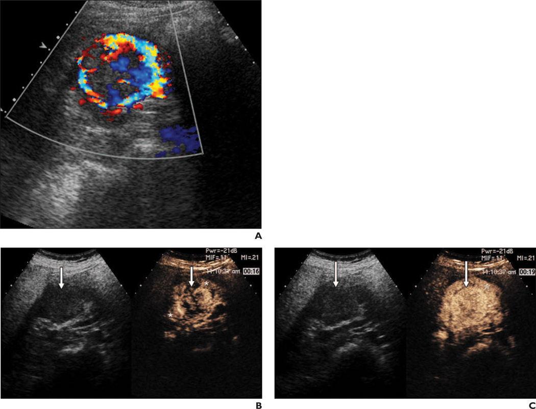
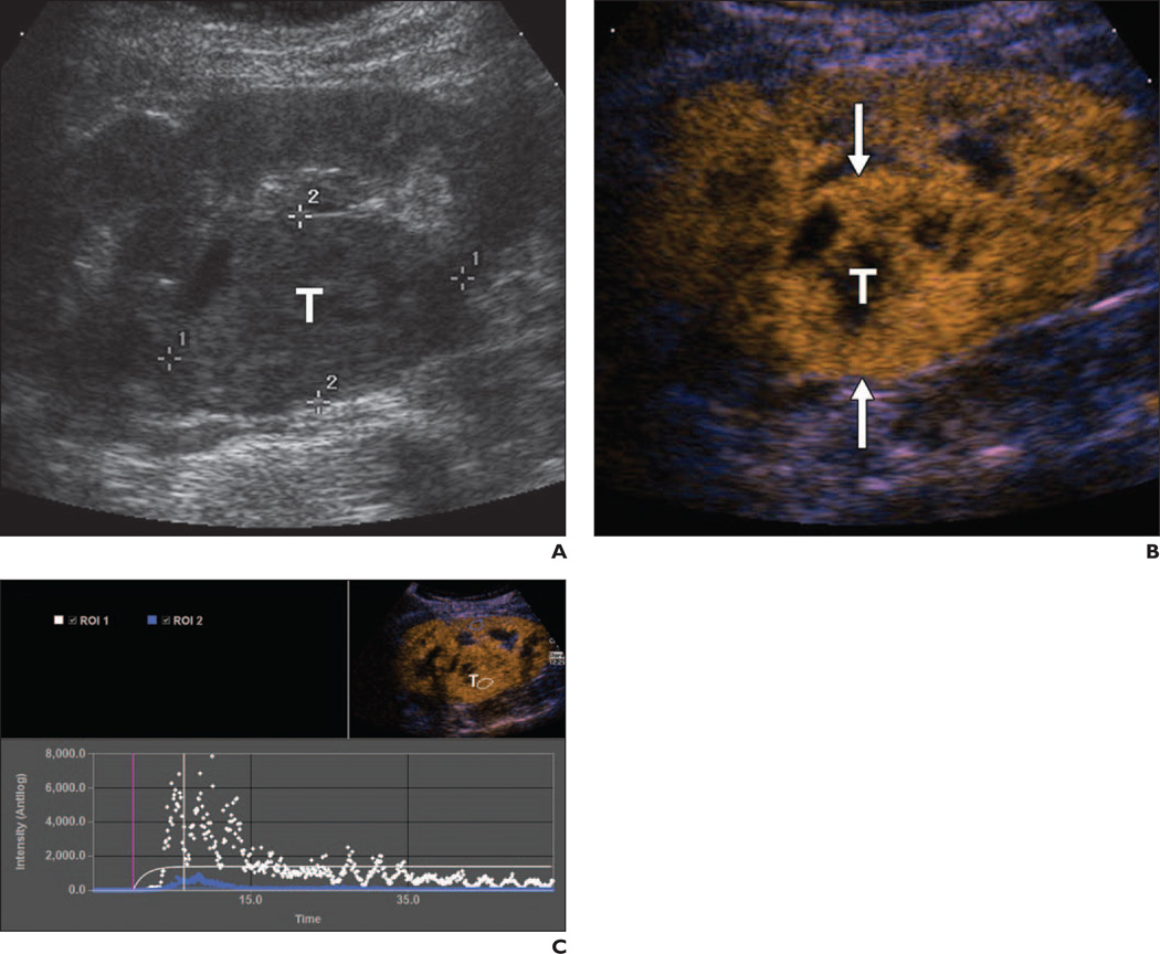
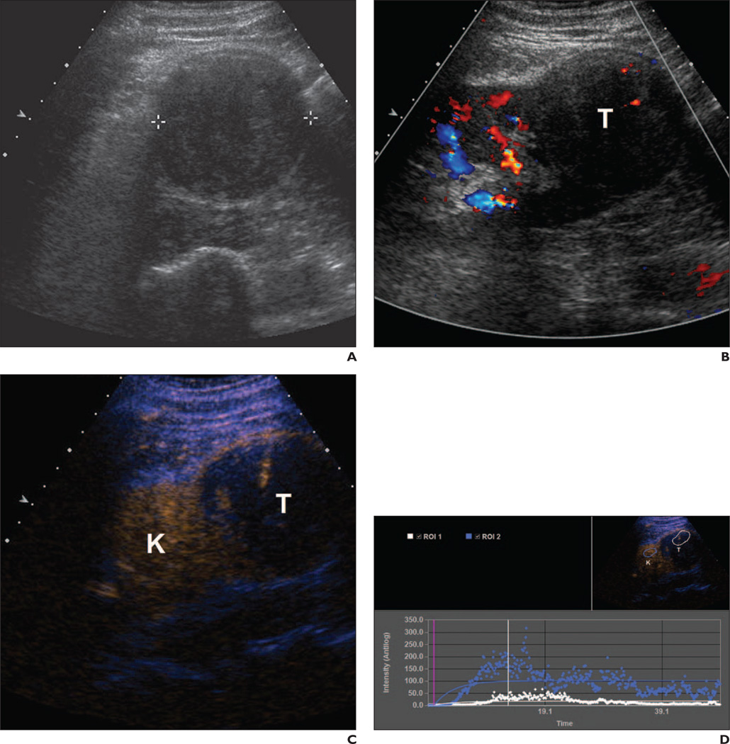
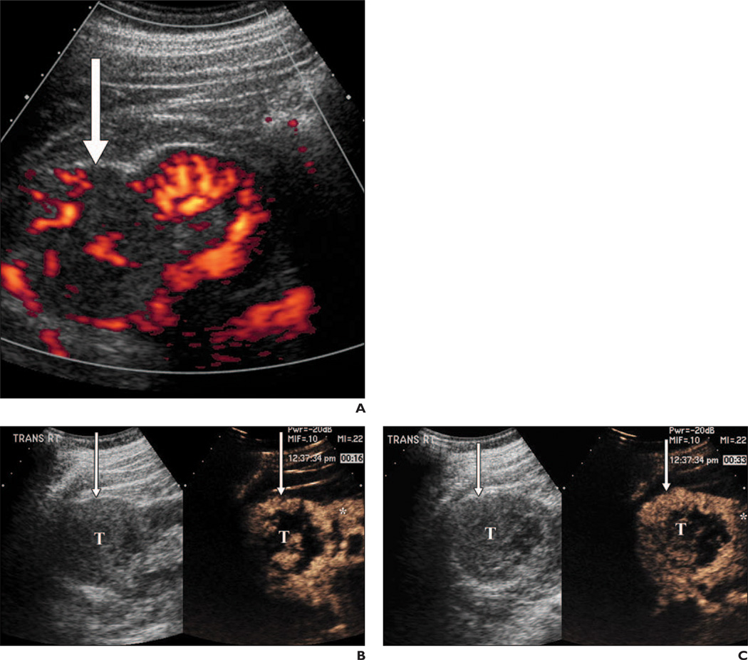
Objective: Nearly 25% of solid renal tumors are indolent cancer or benign and can be managed conservatively in selected patients. This prospective study was performed to determine whether preoperative IV microbubble contrast-enhanced ultrasound can be used to differentiate indolent and benign renal tumors from more aggressive clear cell carcinoma.
Subjects and methods: Thirty-four patients with renal tumors underwent preoperative gray-scale, color, power Doppler, and octafluoropropane microbubble IV contrast-enhanced ultrasound. Three blinded radiologists reading in consensus compared rate of contrast wash-in, grade and pattern of enhancement, and contrast washout compared with adjacent parenchyma. Contrast ultrasound findings were compared with surgical histopathologic findings for all patients.
Results: The 34 patients had 23 clear cell carcinomas, three type 1 papillary carcinomas, one chromophobe carcinoma, one clear rare multilocular low-grade malignant tumor, two unclassified lesions, three oncocytomas, and one benign angiomyolipoma. The combination of heterogeneous lesion echotexture and delayed lesion washout had 85% positive predictive value, 43% negative predictive value, 48% sensitivity, and 82% specificity for predicting whether a lesion was conventional clear cell carcinoma or another tumor. Diminished lesion enhancement grade had 75% positive predictive value, 81% negative predictive value, 55% sensitivity, and 91% specificity for non-clear cell histologic features, either benign or low-grade malignant. Combining delayed washout with quantitative lesion peak intensity of at least 20% of kidney peak intensity had 91% positive predictive value, 40% negative predictive value, 63% sensitivity, and 80% specificity in the prediction of clear cell histologic features.
Conclusion: Ultrasound features of gray-scale heterogeneity, lesion washout, grade of contrast enhancement, and quantitative measure of peak intensity may be useful for differentiating clear cell carcinoma and non-clear cell renal tumors.
Figures
Comment in
-
Re: Evaluation of renal masses with contrast-enhanced ultrasound: initial experience.J Urol. 2012 Apr;187(4):1220. doi: 10.1016/j.juro.2011.12.088. Epub 2012 Feb 14. J Urol. 2012. PMID: 22423900 No abstract available.
References
-
- Reuter VE, Presti JC Jr. Contemporary approach to the classification of renal epithelial tumors. Semin Oncol. 2000;27:124–137. - PubMed
-
- Forman HP, Middleton WD, Melson GL, McClennan BL. Hyperechoic renal cell carcinomas: increase in detection at US. Radiology. 1993;188:431–434. - PubMed
-
- Volpe A, Jewett MA. The role of surveillance for small renal masses. Nat Clin Pract Urol. 2007;4:2–3. - PubMed
-
- Volpe A, Jewett MA. Current role, techniques and outcomes of percutaneous biopsy of renal tumors. Expert Rev Anticancer Ther. 2009;9:773–783. - PubMed
-
- Vasudevan A, Davies RJ, Shannon BA, Cohen RJ. Incidental renal tumours: the frequency of benign lesions and the role of preoperative core biopsy. BJU Int. 2006;97:946–949. - PubMed
Publication types
MeSH terms
Substances
Grants and funding
LinkOut - more resources
Full Text Sources
Medical
