. 2011 Nov;25(11):1849-57.
doi: 10.1210/me.2011-1081. Epub 2011 Sep 22.

Regenerated luminal epithelial cells are derived from preexisting luminal epithelial cells in adult mouse prostate

Affiliations

Regenerated luminal epithelial cells are derived from preexisting luminal epithelial cells in adult mouse prostate

June Liu et al. Mol Endocrinol. 2011 Nov.

Abstract

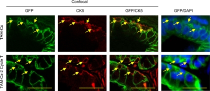
Determining the source of regenerated luminal epithelial cells in the adult prostate during androgen deprivation and replacement will provide insights into the origin of prostate cancer cells and their fate during androgen deprivation therapy. Prostate stem cells in the epithelial layer have been suggested to give rise to luminal epithelium. However, the extent of stem cell participation to prostate regrowth is not clear. In this report, using prostate-specific antigen-CreER(T2)-based genetic lineage marking/tracing in mice, preexisting luminal epithelial cells were shown to be a source of regenerated luminal epithelial cells in the adult prostate. Prostatic luminal epithelial cells could survive androgen deprivation and were capable of proliferating upon androgen replacement. Prostate cancer cells, typically exhibiting a luminal epithelial phenotype, may retain this intrinsic capability to survive and regenerate in response to changes in androgen signaling, providing part of the mechanism for the ultimate failure of androgen deprivation therapy in prostate cancer.


Figures

Fig. 1.
Fig. 1.
Specificity of TAM-inducible PSA-CreERT2 system in mice. A and B, Schematic diagrams showing recombination by active CreERT2 fusion protein and lineage marking of cells in PSA-CreERT2/R26R LacZ and PSA-CreERT2/Rosa26R mT/mG mice. In the presence of TAM, CreERT2 fusion protein causes recombination resulting in removal of the neo or mT gene and permanent, heritable expression of lacZ or GFP, which results in labeling of cells with active PSA promoter. C, TAM-induced lineage marking of luminal epithelial in the lateral prostate. Lineage-marked cells are dark blue via x-gal staining in tissue sections of lateral prostate of 10-wk-old PSA-CreERT2/R26R lacZ or green in PSA-CreERT2/Rosa26R mT/mG mice treated with TAM. Controls are corn oil-injected mice. D, Immunofluorescence staining for CK18 in lateral prostate sections of PSA-CreERT2/Rosa26R mT/mG mice. E, Immunostaining for p63 in lateral prostate sections of PSA-CreERT2/R26R lacZ mice. F, Immunofluorescence staining for CK5 in lateral prostate sections of PSA-CreERT2/Rosa26R mT/mG mice. p63 and CK5 staining are indicated by dashed arrows. Scale bars, 25 μm. mEGFP, membrane-tagged enhanced GFP.
Fig. 2.
Fig. 2.
Androgen dependency of PSA-CreERT2-mediated genetic marking. A, Schematic diagram showing possible outcomes of lineage marking of cells in testis-intact and castrated PSA-CreERT2/R26R LacZ mice. B, TAM induction of genetic marking in testis-intact and castrated PSA-CreERT2/R26R LacZ mice. TAM was injected 3 wk after Cx (Cx-TAM) along with testis-intact control (Intact-TAM) mice. To verify the transient nature of TAM induction, Cx-TAM-2 wk-T and Cx-TAM-4 wk-T groups were generated by implanting testosterone pellets into Cx-TAM mice 2 or 4 wk after the TAM induction. Mice were killed 10 d after TAM injection in Cx-TAM and Intact-TAM groups or 10 d after the testosterone replacement in Cx-TAM-2 wk-T and Cx-TAM-4 wk-T groups. Scale bars, 200 μm. T, Testosterone.
Fig. 3.
Fig. 3.
Survival and regeneration of luminal epithelial cells during cycles of prostate regeneration. A, Predictions from self-duplication model and basal cell model for luminal cell repopulation in regenerating prostate. Regenerated luminal epithelial cells derived from genetically labeled preexisting luminal cells, with either lacZ (blue) or GFP (green), can be determined after regression and regeneration of prostate. Regeneration by self-duplication from preexisting (labeled) cells predicts that the fraction of labeled cells would remain constant in new epithelium. In the basal stem cell model of regeneration, luminal epithelial cells are derived from stem cells that do not express the PSA promoter-driven CreER gene, and the percentage of labeled cells would decrease after each cycle of regression and regeneration. B, Time line employed for TAM labeling and cycles of serial regression and regeneration. C and D, Fate of genetically labeled luminal epithelial cells during cycles of lateral prostate regression and regeneration. Shown are images of x-gal staining and GFP in PSA-CreERT2/R26R LacZ and PSA-CreERT2/Rosa26R mT/mG mouse model, respectively. E and F, Quantitation of the results in C and D, respectively. Number of animals is indicated in parentheses. The percentage of labeled cells was not significantly different between groups in E (P = 0.92) or in F (P = 0.55). Error bars indicate sd. Scale bars, 200 μm. T, Testosterone; Sac, euthanized.
Fig. 4.
Fig. 4.
Proliferation of GFP-labeled luminal epithelial cells after androgen replacement. BrdU was injected 3 d and 4 d after testosterone replacement, and the prostate was isolated 4 h after the second injection (two-cycle group as described in Fig. 3). Red arrows indicate BrdU-labeled, GFP-positive cells (top panel). Green arrows indicate BrdU-negative, GFP-positive cells. Animals without BrdU injection were processed in parallel as negative control. Scale bars, 25 μm. T, Testosterone.
Fig. 5.
Fig. 5.
Confocal microscopic analysis of CK5-stained lateral prostate sections of PSA-CreERT2/Rosa26R mT/mG mice after Cx (TAM-Cx) and two cycles of regeneration (TAM-Cx-2 Cycle T). The lateral prostate sections were from castrated or regenerated prostates as described in Fig 3. Shown are representative images of confocal microscopy of GFP and CK5 staining and regular fluorescent microscopy of GFP and DAPI staining. Arrows indicate CK5-positive basal cells. In confocal images, CK5-positive cells can be surrounded by GFP-positive cells in one focal plane but not in another focal plane. Scale bars, 25 μm. T, Testosterone.

References

    1. Isaacs JT. 1985. Control of cell proliferation and cell death in the normal and neoplastic prostate: a stem cell model. In: Rodgers CH, Coffey DS, Cunha G, Grayhack JT, Henman JR, Horton R. eds. Benign prostatic hyperplasia. Washington DC: Department of Health and Human Services, National Institutes of Health; 85–94
    1. Bonkhoff H, Remberger K. 1996. Differentiation pathways and histogenetic aspects of normal and abnormal prostatic growth: a stem cell model. Prostate 28:98–106 - PubMed
    1. Litvinov IV, Vander Griend DJ, Xu Y, Antony L, Dalrymple SL, Isaacs JT. 2006. Low-calcium serum-free defined medium selects for growth of normal prostatic epithelial stem cells. Cancer Res 66:8598–8607 - PMC - PubMed
    1. Uzgare AR, Xu Y, Isaacs JT. 2004. In vitro culturing and characteristics of transit amplifying epithelial cells from human prostate tissue. J Cell Biochem 91:196–205 - PubMed
    1. Lawson DA, Xin L, Lukacs RU, Cheng D, Witte ON. 2007. Isolation and functional characterization of murine prostate stem cells. Proc Natl Acad Sci USA 104:181–186 - PMC - PubMed

Publication types