. 2011 Oct 1;124(Pt 19):3305-18.
doi: 10.1242/jcs.086934.

Cofilin cooperates with fascin to disassemble filopodial actin filaments

Affiliations

Cofilin cooperates with fascin to disassemble filopodial actin filaments

Dennis Breitsprecher et al. J Cell Sci. .

Abstract

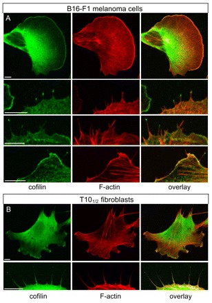
Cells use a large repertoire of proteins to remodel the actin cytoskeleton. Depending on the proteins involved, F-actin is organized in specialized protrusions such as lamellipodia or filopodia, which serve diverse functions in cell migration and sensing. Although factors responsible for directed filament assembly in filopodia have been extensively characterized, the mechanisms of filament disassembly in these structures are mostly unknown. We investigated how the actin-depolymerizing factor cofilin-1 affects the dynamics of fascincrosslinked actin filaments in vitro and in live cells. By multicolor total internal reflection fluorescence microscopy and fluorimetric assays, we found that cofilin-mediated severing is enhanced in fascin-crosslinked bundles compared with isolated filaments, and that fascin and cofilin act synergistically in filament severing. Immunolabeling experiments demonstrated for the first time that besides its known localization in lamellipodia and membrane ruffles, endogenous cofilin can also accumulate in the tips and shafts of filopodia. Live-cell imaging of fluorescently tagged proteins revealed that cofilin is specifically targeted to filopodia upon stalling of protrusion and during their retraction. Subsequent electron tomography established filopodial actin filament and/or bundle fragmentation to precisely correlate with cofilin accumulation. These results identify a new mechanism of filopodium disassembly involving both fascin and cofilin.


Figures

Fig. 1.
Fig. 1.
Dynamics of fascin-mediated actin filament bundling. (A) Polymerization of 1.3 μM actin (30% Alexa-Fluor-488 labeled) in the presence of 500 nM fascin in TIRF buffer. Time is indicated in seconds in the top right of each panel. (B) The fluorescence intensity of the bundle directly correlated with the number of filaments. (C) The number of filaments per bundle formed in the presence of different fascin concentrations. For each condition, at least 30 bundles were analyzed using fluorescence intensity measurements. Boxes indicate 25th percentile, median and 75th percentile of all values; error bars indicate 10th and 90th percentile; minimal and maximal fluorescence intensities are also indicated. The number of filaments per bundle at steady state was only slightly increased at higher fascin concentrations. (D) Time-lapse micrographs of the formation of fascin-crosslinked F-actin bundles. Fascin apparently enhanced the stiffness of the bundles, resulting in kinks and breaks (arrows). All actin filaments within the bundles were oriented in the same direction. Time is indicated in seconds. (E) Time-lapse micrographs of the ‘zippering’ of actin filaments (arrows). The length of the zippered region was measured over time, yielding zippering velocities of ~700 subunits/second. Time is indicated in seconds. Scale bars: 5 μm (A,B,D,E).
Fig. 2.
Fig. 2.
Cofilin-mediated severing of individual filaments and fascincrosslinked bundles. (A) 1.3 μM actin (30% Alexa-Fluor-488 labeled) was polymerized in the presence of 300 nM cofilin in TIRF buffer. The time-lapse micrographs show frequent severing of actin filaments (arrowheads). Time is indicated in seconds. (B) Left: direct visualization of EGFP–cofilin (green) binding to polymerizing Alexa-Fluor-633-labeled actin filaments (30% labeled; red). EGFP–cofilin preferentially bound to and decorated aged actin filaments, resulting in severing (arrowheads), whereas the barbed-end region was neither markedly decorated with EGFP–cofilin nor severed (asterisks). Time is indicated in seconds. Scale bar: 10 μm (A,B). Right: quantification of severing by cofilin and EGFP–cofilin. (C) Cofilin-mediated severing of fascin-bundled filaments. 1.3 μM actin (30% Alexa-Fluor-488 labeled) was polymerized in the presence of 1 μM fascin in TIRF buffer. After 120 seconds, the reaction mixture was replaced with a solution containing 500 nM fascin, 1.3 μM Alexa-Fluor-488–actin and 300 nM cofilin. Although most parts of the fascin-crosslinked filaments were disassembled, short, bundled barbed-end fragments, mainly composed of ATP- and ADP+Pi–actin persisted and continued to grow (arrowheads). Note that the fluorescence intensity of these growing bundles increased over time. Time is indicated in seconds. Scale bar: 5 μm. (D) Histogram of the correlation between cofilin decoration and the length of severed barbed end fragments of single filaments and fascin-crosslinked bundles. The inset shows representative bundled barbed-end fragments after cofilin addition. Values were obtained from experiments equivalent to those shown in A–C. *n=41, #n=44; &n=44; §n=153. (E) Left: as in C, except that 500 nM cofilin was added to the solution. The increase in fluorescence of the actin bundle after cofilin addition was quantified by densitometric analysis of the region in the red box. Right: the plot of fluorescence versus time of the boxed region demonstrates fluorescence increase after cofilin addition (arrow). (F) Cofilin increases the fluorescence of fascin-formed actin bundles in a concentration-dependent manner. The number of filaments per bundle after 10 minutes was estimated by assuming a linear relationship of fluorescence intensity and filament number as shown in Fig. 1B. For each condition, at least 30 bundles were analyzed. Boxes indicate 25th percentile, median and 75th percentile of all values; error bars indicate 10th and 90th percentile; minimal and maximal fluorescence intensities are also indicated. (G) Analyses of the binding of 1 μM cofilin to 1 μM F-actin (15% pyrene labeled) crosslinked by different amounts of fascin in KMEI buffer. Cofilin binding was slowed down by increasing amounts of fascin as shown by the enhanced half-time of the binding reaction. The line is a manual fit of the data points.
Fig. 3.
Fig. 3.
Fascin enhances cofilin severing in a synergistic fashion. (A) Cofilin enhances actin assembly by creating new barbed ends. 4 μM G-actin (10% pyrene labeled) was polymerized in KMEI buffer in the presence of different amounts of cofilin. (B) Fascin enhances cofilin-mediated actin polymerization. 4 μM G-actin (10% pyrene labeled) was polymerized in KMEI buffer in the presence of 600 nM cofilin and increasing amounts of fascin, resulting in a dose-dependent acceleration of actin polymerization. (C) Comparison of polymerization rates mediated by different cofilin concentrations in the presence and absence of fascin. The relative polymerization rates correspond to the maximal slopes of the polymerization reactions shown in A and B. (D) Effects of cofilin and fascin on dilution-induced depolymerization of F-actin. 1 μM F-actin (30% pyrene labeled) either untreated or crosslinked by 0.2 and 1 μM fascin, respectively, was diluted to 100 nM in KMEI buffer with the cofilin concentrations indicated, and spontaneous actin disassembly followed, as measured by pyrene fluorescence. (E) Depolymerization rates of F-actin crosslinked with fascin at the concentrations indicated at different cofilin concentrations. Rates were obtained by measuring the initial slopes of depolymerization reactions as shown in D. (F) Comparison of the enhancement of depolymerization of actin filaments and fascin bundles by cofilin. The fold increase in actin disassembly was obtained by normalizing the depolymerization rates to the respective control experiments without cofilin. (G) Scheme depicting the differential influence of fascin and cofilin on filament depolymerization. (Top) Dilution-induced release of monomers from actin filaments (black arrows) occurs mainly at the barbed end. (Middle) Fascin probably inhibits filament disassembly by crosslinking terminal subunits, thus impairing dissociation of monomers (red arrow). (Bottom) Cofilin antagonizes fascin-mediated resistance to disassembly by creating new filament ends.
Fig. 4.
Fig. 4.
Endogenous cofilin can accumulate in filopodia in different cell types. (A) B16-F1 melanoma cells cultivated on laminin-coated glass coverslips were fixed and labeled with cofilin antibody (green) and filamentous actin with Rhodamine-phalloidin (red). (Top row) Low magnification shows that cofilin accumulates in the entire lamellipodium. (Lower rows) Examples of cofilin enrichment in a subset of filopodia at higher magnification. (B) Subcellular localization of cofilin in T101/2 fibroblasts. (Top row) Cofilin accumulates in actin-rich membrane ruffles at many, individual cellular protrusions. (Bottom row) Cofilin also localizes to the distal tips of filopodial actin filaments in this cell type. The cells were labeled as described for A. All images are three-dimensional reconstructions from confocal sections. Scale bars: 10 μm.
Fig. 5.
Fig. 5.
Cofilin dynamics in protruding and retracting filopodia. (A) mCherry–cofilin expression pattern in a motile B16-F1 cell. During protrusion (upper panels), cofilin only weakly localizes to the base of filopodia and is absent in their tips. Upon retraction (lower panels), cofilin markedly accumulates in the tip region of these structures (arrowheads). Scale bar: 5 μm. (B) Quantification of protruding and retracting filopodia with cofilin accumulated in the tip. Note that cofilin accumulation is exclusively found in retracting filopodia.
Fig. 6.
Fig. 6.
Fascin and cofilin dynamics during protrusion and retraction of filopodia. EGFP–fascin and mCherry–cofilin were coexpressed in B16-F1 cells. Although cofilin is nearly absent in the protruding filopodium, it localizes to the shaft upon cessation of protrusion and accumulates in the tip region upon retraction. Fascin localization in the shaft is prominent during protrusion and decreases upon retraction. Note that fascin can still be found in the tip region of a retracting filopodium. Scale bar: 2 μm. Images were taken at 15-second intervals, and time is given in seconds.
Fig. 7.
Fig. 7.
Comparison of cofilin and actin dynamics during filopodia protrusion and retraction. EGFP–cofilin and mCherry–actin were coexpressed in B16-F1 cells and imaged as indicated. Note the strong actin staining during filopodium protrusion (early time points), and its continuous decrease during cofilin accumulation and cessation of filopodium protrusion and retraction. Scale bar: 3 μm. Images were taken at 15-second intervals, and time is given in seconds.
Fig. 8.
Fig. 8.
Correlated live cell imaging and electron tomography of protruding and retracting filopodia. (A) Time-lapse series (every second frame) of B16-F1 melanoma cell transfected with EGFP–cofilin and mCherry–fascin. Time between the frames shown was 30 seconds. b, c, d indicate individual filopodia that were protruding (b) or retracting (c,d) at the time of fixation after last frame of fluorescence imaging. The final panel shows the fixed cell under phase-contrast optics. Cofilin is present along the retracting filopodium shaft (d) or at the tip (c). (B,C,D) Tomogram sections of the corresponding filopodia in A. The regions indicated by brackets are enlarged in B′,C′ and D′. For technical reasons, the zero tilt image of filopodium ‘c’ is shown in C and C′ (lower panel). Scale bars: 5 μm (A); 250 nm (B–D); 100 nm (B′–D′).
Fig. 9.
Fig. 9.
Proposed mechanism for cofilin-mediated severing of fascincrosslinked filaments. (A) Single actin filaments change their torsional twist upon cofilin (blue circles) binding, resulting in complete decoration of the filament in a cooperative manner. Severing events occur stochastically. Red squares mark sites of severing. (B) Fascin (green bars)-crosslinked filaments are less flexible than free filaments and might therefore be impaired in relaxation upon cofilin binding, resulting in enhanced mechanical stress that leads to enhanced filament severing. Alternatively or in addition, fascin might also impair cooperative binding by cofilin, resulting in a discontinuous decoration of the filament in the bundle and enhanced cofilin severing activity. (C) A model of the potential role of cofilin in filopodium disassembly. During protrusion, filaments are elongated by proteins of the filopodium tip complex (FTP) and immediately crosslinked by fascin, while cofilin is largely absent. Upon cessation of protrusion, cofilin targets to the shaft and strongly accumulates in the tip region of the filopodium by an unknown mechanism, and promotes the disassembly of filopodial actin filaments. Eventually, cofilin-mediated filament severing leads to retraction of the filopodium into the cell body. Upwards and downwards arrow indicates protrusion and retraction, respectively.

References

    1. Adams J. C., Schwartz M. A. (2000). Stimulation of fascin spikes by thrombospondin-1 is mediated by the GTPases Rac and Cdc42. J. Cell Biol. 150, 807-822 - PMC - PubMed
    1. Auinger S., Small J. V. (2008). Correlated light and electron microscopy of the cytoskeleton. Methods in Cell Biol. 88, 257-272 - PubMed
    1. Andrianantoandro E., Pollard T. D. (2006). Mechanism of actin filament turnover by severing and nucleation at different concentrations of ADF/cofilin. Mol. Cell 24, 13-23 - PubMed
    1. Aratyn Y. S., Schaus T. E., Taylor E. W., Borisy G. G. (2007). Intrinsic dynamic behavior of fascin in filopodia. Mol. Biol. Cell 18, 3928-3940 - PMC - PubMed
    1. Bernstein B.W., Bamburg J. R. (2010). ADF/cofilin: a functional node in cell biology. Trends Cell Biol. 4, 187-195 - PMC - PubMed

Publication types