. 2011 Oct 1;124(Pt 19):3344-55.
doi: 10.1242/jcs.087692.

Nuclear relocalisation of cytoplasmic poly(A)-binding proteins PABP1 and PABP4 in response to UV irradiation reveals mRNA-dependent export of metazoan PABPs

Affiliations

Nuclear relocalisation of cytoplasmic poly(A)-binding proteins PABP1 and PABP4 in response to UV irradiation reveals mRNA-dependent export of metazoan PABPs

Hannah M Burgess et al. J Cell Sci. .

Abstract

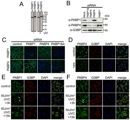
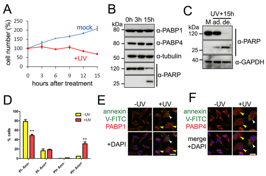
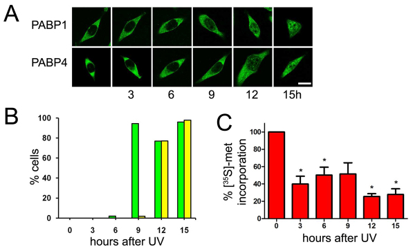
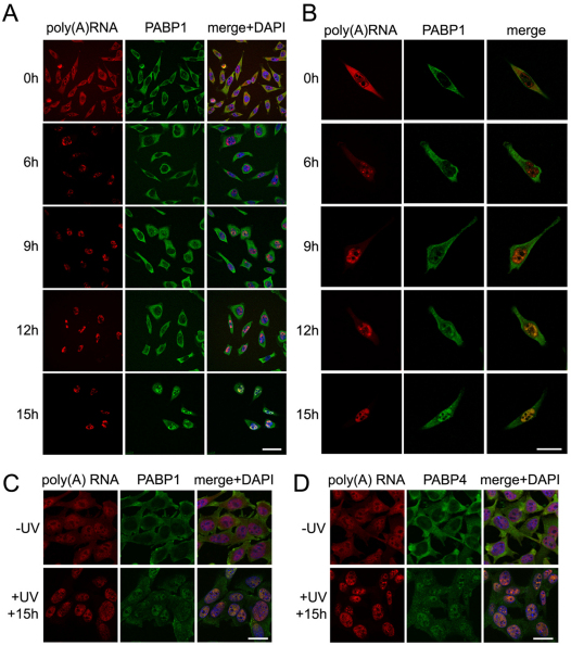
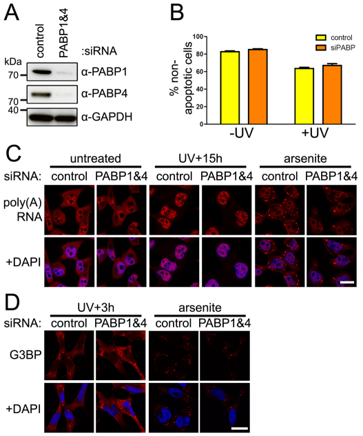
Poly(A)-binding protein 1 (PABP1) has a fundamental role in the regulation of mRNA translation and stability, both of which are crucial for a wide variety of cellular processes. Although generally a diffuse cytoplasmic protein, it can be found in discrete foci such as stress and neuronal granules. Mammals encode several additional cytoplasmic PABPs that remain poorly characterised, and with the exception of PABP4, appear to be restricted in their expression to a small number of cell types. We have found that PABP4, similarly to PABP1, is a diffusely cytoplasmic protein that can be localised to stress granules. However, UV exposure unexpectedly relocalised both proteins to the nucleus. Nuclear relocalisation of PABPs was accompanied by a reduction in protein synthesis but was not linked to apoptosis. In examining the mechanism of PABP relocalisation, we found that it was related to a change in the distribution of poly(A) RNA within cells. Further investigation revealed that this change in RNA distribution was not affected by PABP knockdown but that perturbations that block mRNA export recapitulate PABP relocalisation. Our results support a model in which nuclear export of PABPs is dependent on ongoing mRNA export, and that a block in this process following UV exposure leads to accumulation of cytoplasmic PABPs in the nucleus. These data also provide mechanistic insight into reports that transcriptional inhibitors and expression of certain viral proteins cause relocation of PABP to the nucleus.


Figures

Fig. 1.
Fig. 1.
PABP1 and PABP4 relocalise to SGs early and the nucleus late after UV irradiation. (A) PABP1 and PABP4 antibodies each detect a single protein of the expected size in extracts prepared from untreated HeLa cells or cells 15 hours after treatment with 50 J/m2 UV. (B,C) Specificity of the PABP1 and PABP4 antibodies was demonstrated by western blotting (B) and immunofluorescence (C) following siRNA knockdown in HeLa cells: PABP1 and PABP4 levels were only reduced in response to their respective siRNAs. G3BP served as a loading control. Scale bar: 50 μm. (D) HeLa cells were fixed following exposure to 0.5 mM arsenite treatment or vehicle control (PBS; control) for 1 hour. G3BP (red) and PABP4 (green) were detected by immunofluorescence. (E,F) HeLa cells were fixed at 3 or 15 hours after UV treatment (50 J/m2 254 nm) or 15 hours after mock treatment (control). G3BP (red) and PABP1 or PABP4 (green) were detected by immunofluorescence. Arrowheads indicate UV-induced SGs. Scale bars: 20 μm. In C–F, DNA was stained with DAPI (blue).
Fig. 2.
Fig. 2.
Relocalisation of PABPs is independent of UV-induced apoptosis. (A) Adherent HeLa cells were harvested at the indicated time following treatment with 50 J/m2 UV (red) or mock treatment (blue) and cell numbers were determined by flow cytometry. The number of cells at time zero was set to 100%; values are the means ± s.e.m. of three independent experiments. (B) HeLa cell extracts were prepared from total (adherent and detached) cells 0, 3 or 15 hours after UV treatment (50 J/m2 254 nm) and western blotted with the indicated antibodies. Tubulin was used as a loading control. (C) HeLa cell extracts were prepared from adherent cells 15 hours after mock treatment (M), or from adherent (ad.) or detached (de.) cells 15 hours after UV treatment (50 J/m2, 254 nm) and western blotted with the indicated antibodies. (D) Adherent cells were harvested 15 hours after either UV treatment (50 J/m2 254 nm) or mock treatment and assayed for induction of apoptosis by flow cytometry (supplementary material Fig. S3) using annexin V–FITC and PI staining. Yellow bars: mock treated (−UV) cells; red bars: UV-treated (UV+) cells. Values are means ± s.e.m. of two independent experiments; **P<0.01. (E,F) HeLa cells were fixed 15 hours after UV treatment (50 J/m2 254 nm) or mock treatment and annexin V–FITC (green) was used to stain apoptotic cells, visible as decoration around the cell membrane. PABP1 or PABP4 (red) were detected by immunofluorescence. DNA was stained with DAPI (blue). Fields containing both annexin V-positive (white arrowheads) and -negative (yellow arrowheads) cells were chosen, in which PABP1 (E) or PABP4 (F) was relocalised to the nucleus. Scale bars: 20 μm.
Fig. 3.
Fig. 3.
PABP relocalisation is correlated with decreased protein synthesis. (A) HeLa cells were fixed at 3-hour intervals after UV treatment (50 J/m2 254 nm) or immediately after mock treatment (0 hours) and PABP1 or PABP4 detected by immunofluorescence. Scale bar: 20 μm. (B) The frequency of detectable PABP1 (green bars) or PABP4 (yellow bars) in the nucleus was evaluated in more than 200 cells at each time point. Dividing cells were not counted. Three separate experiments were conducted with very similar results and the data from one such experiment are shown. (C) Protein synthesis rates in HeLa cells at 3-hour intervals after UV treatment (50 J/m2 254 nm) was measured by [35S]methionine incorporation. Each experiment was performed in triplicate and the means normalised to cells assayed immediately after mock treatment (0 hours) and expressed as a percentage. Values are means ± s.e.m. of three independent experiments.
Fig. 4.
Fig. 4.
PABP relocalisation mirrors accumulation of poly(A) RNA in the nucleus after UV treatment in HeLa and NIH3T3 cells. (A,B) HeLa cells were fixed at 3-hour intervals after UV treatment (50 J/m2 254 nm) or after mock treatment (0h). Poly(A) RNA was detected by FISH using a Cy3–oligo(dT)40 probe (red) and PABP1 (green) was detected by immunofluorescence analysis. Single representative cells are shown in B. (C,D) NIH3T3 cells were fixed 15 hours after UV treatment (50 J/m2 254 nm) or mock treatment (−UV) and subject to immunofluorescence analysis as in A. DNA was stained with DAPI (blue). Scale bars: 50 μm (A) and 20 μm (B–D).
Fig. 5.
Fig. 5.
PABP1 and PABP4 are not determinants of poly(A) RNA localisation. (A) Knockdown of PABP1 and PABP4 protein in siRNA-treated HeLa cells was confirmed by western blotting. GAPDH was used as a loading control. (B) After transfection with a control siRNA (yellow bars) or PABP1 and PABP4-specific siRNAs (orange bars), HeLa cells were treated with 50 J/m2 UV followed by 15 hours incubation, and adherent cells assayed for annexin V–FITC and PI staining by flow cytometry. Percentage annexin V-negative and PI-negative cells are shown. Data represented as means ± s.e.m. of three independent experiments. (C) Control or PABP1 and PABP4 siRNA-transfected HeLa cells were treated with 50 J/m2 UV followed by 15 hours incubation or with 0.5 mM sodium arsenite for 1 hour. Poly(A) RNA was detected by FISH using a Cy3–oligo(dT)40 probe (red). DNA was stained by DAPI (blue). Scale bar: 20 μm. (D) siRNA-transfected HeLa cells were treated with 50 J/m2 UV followed by 3 hours incubation or with 0.5 mM sodium arsenite for 1 hour. The cells were then fixed and G3BP (red) detected by immunofluorescence. DNA was stained with DAPI (blue). Scale bar: 20 μm.
Fig. 6.
Fig. 6.
PABP relocalises to the nucleus when mRNA export is blocked. (A) HeLa cells were transfected with 350 ng/ml ICP27–GFP. 24 hours post-transfection, poly(A) RNA (top panel) was detected by FISH (red) and PABP1 and PABP4 (middle and bottom panels, respectively) detected by immunofluorescence (red). (B) HeLa cells were transfected with 3 μg shTAP or control plasmid and 1 μg GFP (pEGFP-C1). A cell transfected with shTAP is indicated by an arrowhead. 65 hours post-transfection poly(A) RNA was detected by FISH using a Cy3–oligo(dT)40 probe (red). (C,D) HeLa cells were transfected with 3 μg shTAP or control plasmid and 65 hours post-transfection TAP (red) and PABP1 (C, green) or PABP4 (D, green) were detected by immunofluorescence. Arrowheads indicate cells in which TAP knockdown was efficient. DNA was stained with DAPI (blue). Scale bars: 20 μm.

Similar articles

Cited by

References

    1. Afonina E., Stauber R., Pavlakis G. N. (1998). The human poly(A)-binding protein 1 shuttles between the nucleus and the cytoplasm. J. Biol. Chem. 273, 13015-13021 - PubMed
    1. Albrecht M., Lengauer T. (2004). Survey on the PABC recognition motif PAM2. Biochem. Biophys. Res. Commun. 316, 129-138 - PubMed
    1. Anderson P., Kedersha N. (2009). RNA granules: post-transcriptional and epigenetic modulators of gene expression. Nat. Rev. Mol. Cell Biol. 10, 430-436 - PubMed
    1. Apponi L. H., Leung S. W., Williams K. R., Valentini S. R., Corbett A. H., Pavlath G. K. (2010). Loss of nuclear poly(A)-binding protein 1 causes defects in myogenesis and mRNA biogenesis. Hum. Mol. Genet. 19, 1058-1065 - PMC - PubMed
    1. Batista L. F., Kaina B., Meneghini R., Menck C. F. (2009). How DNA lesions are turned into powerful killing structures: insights from UV-induced apoptosis. Mutat. Res. 681, 197-208 - PubMed

Publication types

MeSH terms