

MR angiography and imaging for the evaluation of middle cerebral artery atherosclerotic disease
- PMID: 21940802
- PMCID: PMC7966534
- DOI: 10.3174/ajnr.A2697
MR angiography and imaging for the evaluation of middle cerebral artery atherosclerotic disease
Abstract
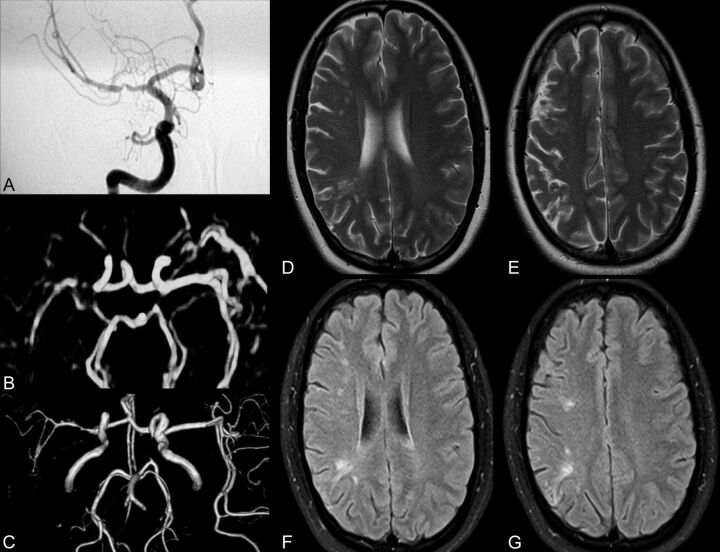
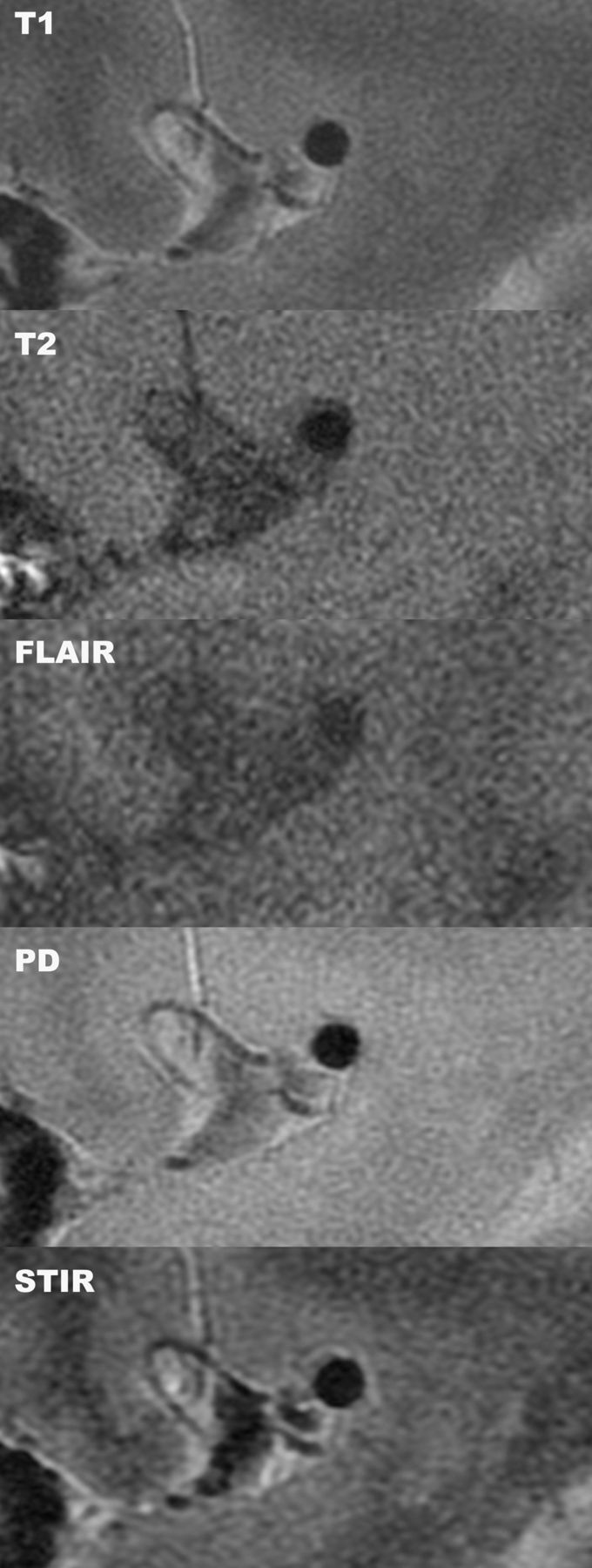
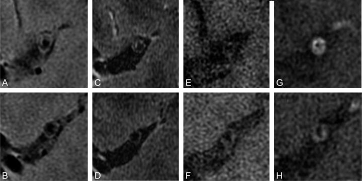
Intracranial atherosclerotic disease may constitute the most common cause of ischemic stroke worldwide; yet, in the developed world, imaging research has largely focused on extracranial atherosclerosis. Many studies in populations of Asian, African, and Hispanic descent demonstrate the preponderance of intracranial stenosis compared with carotid stenosis. This review examines the clinical presentations of MCA atherosclerosis and stenosis and the use of noninvasive MR imaging in the assessment of intracranial vasculature. MRA is a well-validated technique that offers great advantage over traditional angiography. Advances in high-resolution MR imaging of MCA stenosis have the potential to yield excellent visualization of plaque. Future developments in high-resolution MR imaging to depict intracranial atherosclerosis are explored in this review; these advances will guide endovascular therapy and the comparison of novel interventions.
Figures
References
-
- World Health Organization. Causes of Death. Geneva, Switzerland: World Health Organization; 2004. http://www.who.int/healthinfo/global_burden_disease/GBD_report_2004updat.... Accessed September 15, 2011.
-
- Sridharan SE, Unnikrishnan JP, Sukumaran S, et al. . Incidence, types, risk factors, and outcome of stroke in a developing country: the Trivandrum Stroke Registry. Stroke 2009; 40: 1212– 18 - PubMed
-
- Arenillas JF. Intracranial atherosclerosis: current concepts. Stroke 2011; 42 (1 suppl): S20– 23 - PubMed
-
- Lhermitte F, Gautier JC, Derouesne C, et al. . Ischemic accidents in the middle cerebral artery territory: a study of the causes in 122 cases. Arch Neurol 1968; 19: 248– 56 - PubMed
-
- Suwanwela NC, Chutinetr A. Risk factors for atherosclerosis of cervicocerebral arteries: intracranial versus extracranial. Neuroepidemiology 2003; 22: 37– 40 - PubMed
Publication types
MeSH terms
LinkOut - more resources
Full Text Sources