. 2011 Dec;31(12):e100-6.
doi: 10.1161/ATVBAHA.111.238261. Epub 2011 Sep 22.

Protease-activated receptor-2 modulates protease-activated receptor-1-driven neointimal hyperplasia

Affiliations

Protease-activated receptor-2 modulates protease-activated receptor-1-driven neointimal hyperplasia

Leila M Sevigny et al. Arterioscler Thromb Vasc Biol. 2011 Dec.

Abstract

Objective: Emerging evidence suggests that protease-activated receptors-1 and -2 (PAR1 and PAR2) can signal together in response to proteases found in the rapidly changing microenvironment of damaged blood vessels. However, it is unknown whether PAR1 and PAR2 promote or mitigate the hyperplastic response to arterial injury. Using cell-penetrating PAR1 pepducins and mice deficient in PAR1 or PAR2, we set out to determine the respective contributions of the receptors to hyperplasia and phenotypic modulation of smooth muscle cells (SMCs) in response to arterial injury.

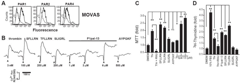
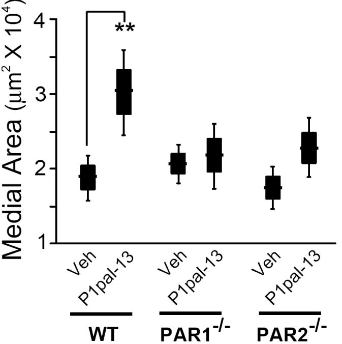
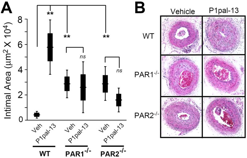
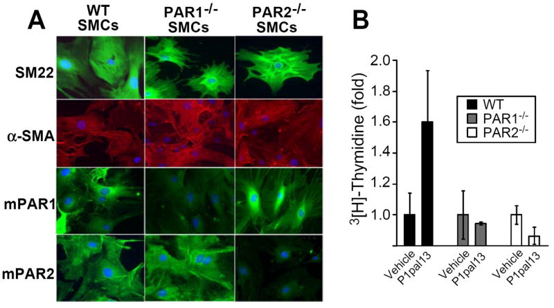
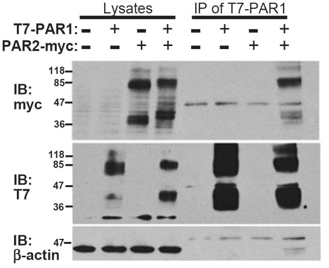
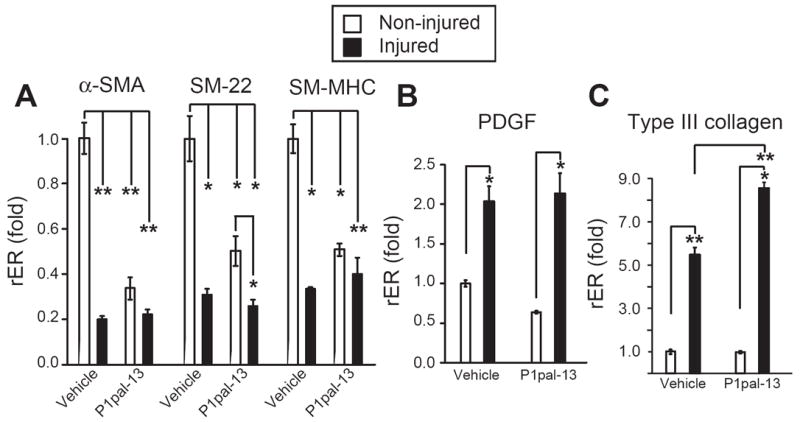
Methods and results: SMCs were strongly activated by PAR1 stimulation, as evidenced by increased mitogenesis, mitochondrial activity, and calcium mobilization. The effects of chronic PAR1 stimulation following vascular injury were studied by performing carotid artery ligations in mice treated with the PAR1 agonist pepducin, P1pal-13. Histological analysis revealed that PAR1 stimulation caused striking hyperplasia, which was ablated in PAR1(-/-) and, surprisingly, PAR2(-/-) mice. P1pal-13 treatment yielded an expression pattern consistent with a dedifferentiated phenotype in carotid artery SMCs. Detection of PAR1-PAR2 complexes provided an explanation for the hyperplastic effects of the PAR1 agonist requiring the presence of both receptors.

Conclusions: We conclude that PAR2 regulates the PAR1 hyperplastic response to arterial injury leading to stenosis.


Figures

Figure 1
Figure 1. PAR1 agonists stimulate calcium mobilization and proliferation of smooth muscle cells (SMCs)
(A) Mouse vascular aorta SMCs (MOVAS) were analyzed for surface expression of PAR1, PAR2, and PAR4 by flow cytometry. (B) Profile of PAR agonist activity in mobilizing calcium in MOVAS, (C) in mitochondrial activity as assessed by MTT (0.3 nM thrombin, 3 μM RWJ-56110, 100 μM SFLLRN, TFLLRN, or SLIGRL, 200 μM AYPGKF, or P1pal-13 as indicated for 4 d), (D) and in mitogenesis assays as assessed by 3[H]-Thymidine (P1pal-13 was used at 3 μM, other agonists and inhibitors were used at the same concentrations in C, for 2 d).*, P <0.05 and **, P <0.005.
Figure 2
Figure 2. The PAR1 pepducin agonist, P1pal-13, causes a significant increase in medial hyperplasia that is lost in PAR1- and PAR2-deficient mice
C57BL/6 wild-type (WT, n=12-17), PAR1-/- (n=7-10), and PAR2-/- (n=9-19) mice underwent ligation injury of the left common carotid artery and were treated daily for 21 d with P1pal-13 (2.5 mg/kg) or vehicle (20% DMSO). The mean of each treatment group is indicated as a horizontal line within a black box that depicts ± se and the error bars depict ± 2 se (to represent the 95% confidence interval) for each treatment cohort calculated from cross sections of the arteries as described in the Supplemental Material. **, P <0.005.
Figure 3
Figure 3. The PAR1 pepducin agonist causes significant neointimal hyperplasia in the injured carotid arteries of wild-type but not PAR1- or PAR2-deficient mice
C57BL/6 wild-type (WT, n=12-17), PAR1-/- (n=7-10), and PAR2-/- (n=9-19) underwent ligation injury of the left common carotid artery and were treated as described in Figure 2. (A) The mean intimal areas (black boxes represent ± se and error bars represent ± 2se) for each treatment cohort were calculated from cross sections of the arteries as described in the Supplemental Material. **, P <0.005. (B) Representative H&E cross-sections of carotids from different treatment groups and genotypes.
Figure 4
Figure 4. Effect of genetic loss of PAR1 or PAR2 on proliferation
(A) Primary SMCs were isolated from the carotid arteries of wild-type (WT), PAR1-/- and PAR2-/- mice. SMCs were plated on culture slides and stained with DAPI (blue), or antibodies against SM-22 (top row, green/FITC), α-SMA (second row, red/TRITC), mouse PAR1 (third row, green/FITC), or mouse PAR2 (bottom row, green/FITC). (B) Primary SMCs were subjected to daily treatment of vehicle (0.2% DMSO) or P1pal-13 (3 μM) for 3 d and mitogenesis was measured by incorporation of 3[H]-Thymidine.
Figure 5
Figure 5. Co-immunoprecipitation of PAR1 with PAR2
(top) T7-PAR1 was transiently co-expressed in COS7 cells with PAR2-myc or pcDEF3 vector alone (-) as indicated. T7 agarose beads were used to immunoprecipitate bound PAR2-myc from cell lysates as shown by the myc immunoblot. (middle) Immunoblot of T7 confirmed the presence of T7-PAR1, (bottom) and β-actin staining confirmed equal loading of the lysates.
Figure 6
Figure 6. The PAR1 agonist pepducin causes de-differentiation of SMCs isolated from the carotid arteries of mice
(A) RNA was extracted from uninjured and injured (ligated) carotid arteries of C57BL/6 wild-type mice treated with P1pal-13 or vehicle for 14 days. Real-time PCR was performed with primers for markers of SMC differentiation, (B,C) or markers of SMC de-differentiation. Mean relative gene expression was normalized to GAPDH expression (rER, relative expression ratio) and fold change was determined by comparing the relative expression ratios (rER) of each group to uninjured vehicle treated arteries (n=3), *, P <0.05 and **, P <0.005.

Comment in

References

    1. Orford JL, Selwyn AP, Ganz P, Popma JJ, Rogers C. The comparative pathobiology of atherosclerosis and restenosis. Am J Cardiol. 2000;86:6H–11H. - PubMed
    1. Douglas JS., Jr Pharmacologic approaches to restenosis prevention. Am J Cardiol. 2007;100:10K–16K. - PubMed
    1. Sukhija R, Mehta JL, Sachdeva R. Present status of coronary bifurcation stenting. Clin Cardiol. 2008;31:63–66. - PMC - PubMed
    1. Leger AJ, Covic L, Kuliopulos A. Protease-activated receptors in cardiovascular diseases. Circulation. 2006;114:1070–1077. - PubMed
    1. O’Brien PJ, Molino M, Kahn M, Brass LF. Protease activated receptors: theme and variations. Oncogene. 2001;20:1570–1581. - PubMed

Publication types

MeSH terms