Review
. 2011 Sep 23;11(10):693-702.
doi: 10.1038/nri3063.

Highlights of 10 years of immunology in Nature Reviews Immunology

Affiliations
Review

Highlights of 10 years of immunology in Nature Reviews Immunology

Ruslan Medzhitov et al. Nat Rev Immunol. .

Abstract

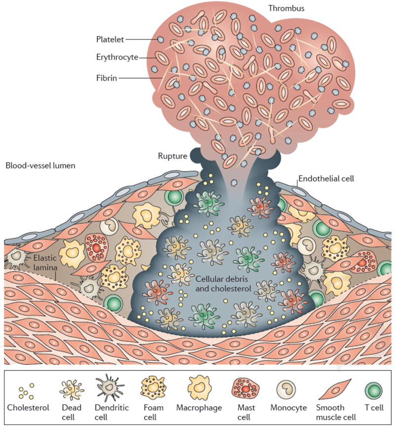
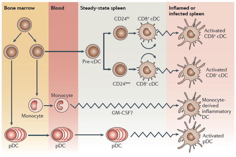
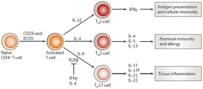
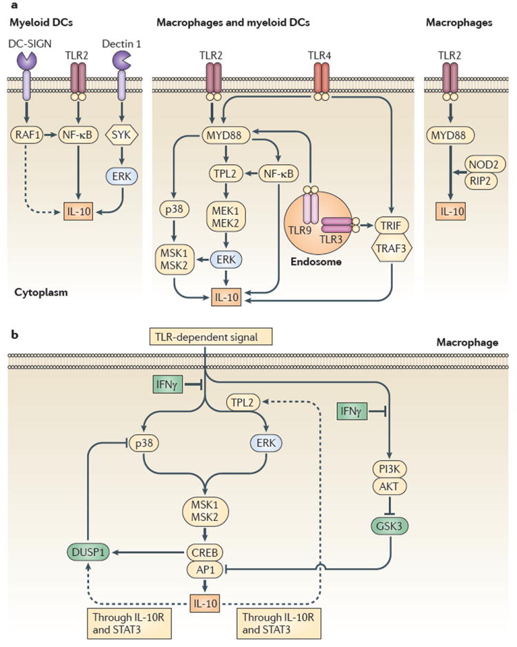
As Nature Reviews Immunology reaches its 10(th) anniversary, the authors of one of the top-cited articles from each year take a trip down memory lane. We've asked them to look back on the state of research at the time their Review was published, to consider why the article has had the impact it has and to discuss the future directions of their field. This Viewpoint article provides an interesting snapshot of some of the fundamental advances in immunology over the past 10 years. Highlights include our improved understanding of Toll-like receptor signalling, and of immune regulation mediated by regulatory T cells, indoleamine 2,3-dioxygenase, myeloid-derived suppressor cells and interleukin-10. The complexities in the development and heterogeneity of macrophages, dendritic cells and T helper cells continue to engage immunologists, as do the immune processes involved in diseases such as atherosclerosis. We look forward to what the next 10 years of immunology research may bring.


Figures

Figure 1
Figure 1. Toll signalling pathways
The Toll-like receptor (TLR) and interleukin-1 receptor (IL-1R)-family members share several signalling components, including the adaptor MyD88, Toll-interacting protein (TOLLIP), the protein kinase IRAK (IL-1R-associated kinase) and TRAF6 (TNF receptor-associated factor 6). TRAF6 can activate nuclear factor-κB (NF-κB) through TAK1 (TGF-β-activated kinase), and JNK (c-Jun N-terminal kinase) and p38 MAP kinases through MKK6 (mitogen-activated protein kinase kinase 6). TLR4 signals through another adaptor in addition to MyD88–TIRAP (Toll/interleukin-1 (IL-1) receptor domain-containing adaptor protein), which activates MyD88-independent signalling downstream of TLR4. The protein kinase PKR functions downstream of TIRAP, but its importance in this pathway has not yet been established. Image is reproduced, with permission, from REF. © (2001) Macmillan Publishers Ltd. All rights reserved.
Figure 2
Figure 2. Plaque activation, rupture and thrombosis
When activated, immune cells including macrophages, T cells and mast cells can release pro-inflammatory cytokines, which reduce collagen formation and induce the expression of tissue factor. Proteases that attack the collagenous cap are also released by activated immune cells. The weakened plaque might fissure when subjected to the forces of arterial blood pressure. Exposure of subendothelial structures and procoagulants such as tissue factor promotes platelet aggregation and thrombosis. A thrombus forms and might occlude the lumen of the artery, leading to acute ischaemia. Image is reproduced, with permission, from REF. © (2006) Macmillan Publishers Ltd. All rights reserved.
Figure 3
Figure 3. Pathways to splenic dendritic cells
Many, branching pathways are involved in generating the dendritic cells (DCs) found in the spleen. The conventional DCs (cDCs) in the spleen of steady-state mice derive from an intrasplenic precursor, a pre-cDC. This precursor population might be replenished from earlier precursor cells that are generated in the bone marrow, which might occasionally seed the spleen from the bloodstream. Alternatively, as the spleen remains a haematopoietic organ in mice, the pre-cDCs might be generated endogenously. A late branch in the cDC developmental pathway, detected by high or low expression of CD24 on the precursor cells, leads to pre-cDCs in the spleen that are pre-committed to form either CD8+ or CD8- cDCs, respectively. The cDCs so formed are in an immature state and are still capable of some homeostatic proliferation. In contrast to the cDCs, the plasmacytoid DCs (pDCs) are generated in the bone marrow by a pathway that branches off from that of cDCs. The pDCs found in the mouse spleen and other tissues probably arrive there from the bloodstream. This steady-state situation changes after microbial stimulation or inflammation. In addition to full activation of the resident cDCs and the pDCs in the spleen, a new type of ‘inflammatory DC’ is then generated from monocytes, a DC type that is not present in the steady state. GM-CSF, granulocyte/macrophage colony-stimulating factor. Image is reproduced, with permission, from REF. © (2007) Macmillan Publishers Ltd. All rights reserved.
Figure 4
Figure 4. General scheme of T-helper-cell differentiation
Naive CD4+ T cells, after activation by signalling through the T-cell receptor and co-stimulatory molecules such as CD28 and inducible T-cell co-stimulator (ICOS), can differentiate into one of three lineages of effector T helper (TH) cells — TH1, TH2 or TH17 cells. These cells produce different cytokines and have distinct immunoregulatory functions. Interferon-γ (IFNγ) produced by TH1 cells is important in the regulation of antigen presentation and cellular immunity. The TH2-cell cytokines interleukin-4 (IL-4), IL-5 and IL-13 regulate B-cell responses and anti-parasite immunity and are crucial mediators of allergic diseases. TH17 cells have been shown to express IL-17, IL-17F, IL-21 and IL-22 (and IL-26 in humans) and to regulate inflammatory responses. TGFβ, transforming growth factor-β. Image is reproduced, with permission, from REF. © (2008) Macmillan Publishers Ltd. All rights reserved.
Figure 5
Figure 5. Signals that induce interleukin-10 expression by cells of the innate immune response
a ∣ The expression of interleukin-10 (IL-10) can be induced by Toll-like receptor (TLR) or non-TLR signalling in macrophages and myeloid dendritic cells (DCs). Activation of TLRs and their adaptor molecules — myeloid differentiation primary-response protein 88 (MYD88) and TIR-domain-containing adaptor protein inducing IFNβ (TRIF) — results in the activation of the extracellular signal-regulated kinase 1 (ERK1) and ERK2 (which are collectively referred to here as ERK), p38 and nuclear factor-κB (NF-κB) pathways. Activation of these pathways results in the induction of IL-10 expression, in addition to pro-inflammatory cytokines. In myeloid DCs, non-TLR signals through DC-specific ICAM3-grabbing non-integrin (DC-SIGN) and RAF1 can augment TLR2-induced IL-10 production. Furthermore, activation of dectin 1 and the signalling molecules spleen tyrosine kinase (SYK) and ERK results in IL-10 production. In macrophages, a role for nucleotide-binding oligomerization domain 2 (NOD2) signalling in IL-10 induction, in crosstalk with TLR2, has been described. b ∣ Positive and negative feedback loops for IL-10 regulation in macrophages. The p38 and ERK pathways leading to IL-10 expression by macrophages are tightly controlled by interferon-γ (IFNγ) and IL-10 itself. IL-10 feeds back to induce the expression of dual-specificity protein phosphatase 1 (DUSP1), which negatively regulates p38 phosphorylation and thus limits IL-10 production. IL-10 can also positively feed back to upregulate tumour progression locus 2 (TPL2) expression, thus providing a positive amplification loop for its own production. In addition, IFNγ can also interfere with the phosphoinositide 3-kinase (PI3K)–AKT pathway, releasing glycogen synthase kinase 3 (GSK3). As GSK3 normally blocks IL-10 expression by acting on the transcription factors cAMP response element-binding protein (CREB) and activator protein 1 (AP1), IL-10 production is inhibited by IFNγ through its effects on PI3K. IL-10R, IL-10 receptor; MEK, MAPK/ERK1 kinase; MSK, mitogen-and stress-activated protein kinase; RIP2, receptor-interacting protein 2; STAT3, signal transducer and activator of transcription 3; TRAF3, TNFR-associated factor 3. Image is reproduced, with permission, from REF. © (2010) Macmillan Publishers Ltd. All rights reserved.

References

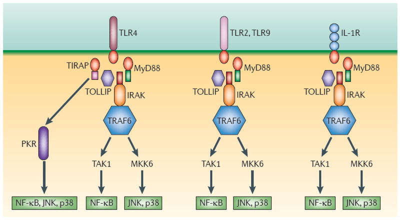
    1. Medzhitov R. Toll-like receptors and innate immunity. Nature Rev Immunol. 2001;1:135–145. - PubMed
    1. Shevach E. CD4+CD25+ suppressor T cells: more questions than answers. Nature Rev Immunol. 2002;2:389–400. - PubMed
    1. Sakaguchi S, Sakaguchi N, Asano M, Itoh M, Toda M. Immunologic self-tolerance maintained by activated T cells expressing IL-2 receptor α-chains (CD25) J Immunol. 1995;155:1151–1164. - PubMed
    1. Shevach EM. Certified professionals: CD4+CD25+ suppressor T cells. J Exp Med. 2001;193:F41–F45. - PMC - PubMed
    1. Horri S, Nomura T, Sakaguchi S. Control of T regulatory cell development by the transcription factor Foxp3. Science. 2003;299:1057–1062. - PubMed

MeSH terms