Review
. 2011 Dec;18(12):1815-24.
doi: 10.1038/cdd.2011.120. Epub 2011 Sep 23.

Biological functions of p53 isoforms through evolution: lessons from animal and cellular models

Affiliations
Review

Biological functions of p53 isoforms through evolution: lessons from animal and cellular models

V Marcel et al. Cell Death Differ. 2011 Dec.

Abstract

The TP53 tumour-suppressor gene is expressed as several protein isoforms generated by different mechanisms, including use of alternative promoters, splicing sites and translational initiation sites, that are conserved through evolution and within the TP53 homologues, TP63 and TP73. Although first described in the eighties, the importance of p53 isoforms in regulating the suppressive functions of p53 has only become evident in the last 10 years, by analogy with observations that p63 and p73 isoforms appeared indispensable to fully understand the biological functions of TP63 and TP73. This review summarizes recent advances in the field of 'p53 isoforms', including new data on p63 and p73 isoforms. Details of the alternative mechanisms that produce p53 isoforms and cis- and trans-regulators identified are provided. The main focus is on their biological functions (apoptosis, cell cycle, aging and so on) in cellular and animal models, including mouse, zebrafish and Drosophila. Finally, the deregulation of p53 isoform expression in human cancers is reviewed. Based on these latest results, several developments are expected in the future: the identification of drugs modulating p53 isoform expression; the generation of animal models and the evaluation of the use of p53 isoform as biomarkers in human cancers.


Figures
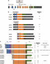
Figure 1
Figure 1
A schematic representation of human p53 isoforms. (a) The human TP53 gene structure. The TP53 gene, which consists of 11 exons (coloured boxes, coding exons; grey boxes, non-coding exons), expresses several p53 isoforms owing to usage of alternative promoters (↱), splicing sites (^) or translational initiation sites (∣). (b) Human p53 mRNA variants. The proximal promoter P1, located upstream from exon-1, regulates the transcription of two transcripts: the fully spliced p53 mRNA (FSp53), which encodes both p53 (from ATG1) and Δ40p53 forms (from ATG40), and the p53I2 mRNA, retaining the entire intron-2 by alternative splicing, which generates Δ40p53 forms from ATG40, owing to the presence of stop codons (*) in the reading frame starting from ATG1. The internal P2 promoter, described as encompassing the region from intron-1 to exon-5, produces p53I4 mRNA, initiated in intron-4 and encoding the N-terminal Δ133p53 (from ATG133) and Δ160p53 forms (from ATG160). Three different C-terminal p53 forms have been described owing to alternative splicing of intron-9: the α-forms resulting from the excision of the entire intron-9, and the β- and γ-forms produced by retention of two small parts of intron-9. Some cis- and trans-regulators driving p53 isoform expression have been described (purple boxes). Endogenous expression of most of the p53 mRNA variants in human cells has been reported (references shown in parentheses). Grey box, non-coding sequence; NR, not yet reported. (c) Human p53 protein isoforms. The canonical p53 protein contains a TAD (blue), a proline-rich domain (PXXP, purple), a DNA-binding domain (DBD, orange) and an OD (green) that encompasses a nuclear localization domain (NLS, green) and five regions conserved through evolution (I–V in grey boxes). Compared with p53, the Δ40p53 forms lack the first TAD, whereas the Δ133p53 and Δ160p53 isoforms lack the entire TAD and parts of the DBD. At the C-terminal, the α-peptide corresponds to the OD that is replace by new residues, the β- and γ-peptides (brown). On the right is indicated the theoretical molecular weight, the detection at endogenous levels (reference in brackets) as well as the different names of the isoforms used in the literature. The color reproduction of this figure is available at the Cell Death and Differentiation journal online
Figure 2
Figure 2
p53 isoforms in animal models. (a) Structural organization of p53 isoforms through evolution. Like humans, mouse, Drosophila and zebrafish express a full-length p53 protein, which conserves a TAD (blue), a DNA-binding domain (DBD, orange) and an OD (green). Only the mouse Mp53 protein presents a proline-rich domain (PXXP, purple) and a nuclear localization signal (NLS, green). In addition, all these animals express some p53 isoforms that have the same structural organization as the human p53 isoforms owing to the use of alternative promoters and splicing sites. M, mouse protein; D, Drosophila protein; Z, zebrafish protein; N-terminal p53 isoform identification, Δ forms; N-terminal p53 isoform denomination, codon number, when initiated ATG occurs in the coding sequence, or N, when initiation occurs in a non-coding sequence; C-terminal p53 isoform identification, AS (alternative splicing, green boxes); grey box, different residues compared with the full-length p53 protein. (b) Localization of translation initiation sites in animal p53 sequences. Red, ATG1 generating the full-length p53 protein; green, the methionine used to produce the homologues to the human Δ40p53 forms; blue, the methionine used to produce the homologues to the human Δ133p53 forms; orange, the methionine used to produce the homologues to the human Δ160p53 forms. The color reproduction of this figure is available at the Cell Death and Differentiation journal online
Figure 3
Figure 3
p53 isoforms in the p53 network. p53 integrates the different stress signals to adapt cell fate to the intensity and the nature of stress by regulating several biological functions to maintain genomic and cellular integrity. In addition, p53 controls physiological functions under basal conditions. Recent data suggest that p53 isoforms modulate p53-mediated cell fate outcome and may thus be key components of the p53-mediated decision not only in response to stress but also under basal conditions. It has also been reported that p53 isoforms have p53-independent activities and directly regulate cell-cycle arrest and apoptosis. In addition, genetic alterations, such as TP53 SNPs and TP53 mutations, affect the expression of p53 isoforms, which may result in tumorigenesis

References

    1. Levine AJ, Oren M. The first 30 years of p53: growing ever more complex. Nat Rev Cancer. 2009;9:749–758. - PMC - PubMed
    1. Levine AJ, Tomasini R, McKeon FD, Mak TW, Melino G. The p53 family: guardians of maternal reproduction. Nat Rev Mol Cell Biol. 2011;12:259–265. - PubMed
    1. Meek DW, Anderson CW. Posttranslational modification of p53: cooperative integrators of function. Cold Spring Harb Perspect Biol. 2009;1:a000950. - PMC - PubMed
    1. Vilborg A, Wilhelm MT, Wiman KG. Regulation of tumor suppressor p53 at the RNA level. J Mol Med. 2010;88:645–652. - PubMed
    1. Garritano S, Gemignani F, Palmero EI, Olivier M, Martel-Planche G, Le Calvez-Kelm F, et al. Detailed haplotype analysis at the TP53 locus in p.R337H mutation carriers in the population of Southern Brazil: evidence for a founder effect. Hum Mutat. 2010;31:143–150. - PubMed

Publication types

MeSH terms

Substances

LinkOut - more resources