. 2011 Sep 7:5:59.
doi: 10.3389/fnana.2011.00059. eCollection 2011.

Basal Ganglia disorders associated with imbalances in the striatal striosome and matrix compartments

Affiliations

Basal Ganglia disorders associated with imbalances in the striatal striosome and matrix compartments

Jill R Crittenden et al. Front Neuroanat. .

Abstract

The striatum is composed principally of GABAergic, medium spiny striatal projection neurons (MSNs) that can be categorized based on their gene expression, electrophysiological profiles, and input-output circuits. Major subdivisions of MSN populations include (1) those in ventromedial and dorsolateral striatal regions, (2) those giving rise to the direct and indirect pathways, and (3) those that lie in the striosome and matrix compartments. The first two classificatory schemes have enabled advances in understanding of how basal ganglia circuits contribute to disease. However, despite the large number of molecules that are differentially expressed in the striosomes or the extra-striosomal matrix, and the evidence that these compartments have different input-output connections, our understanding of how this compartmentalization contributes to striatal function is still not clear. A broad view is that the matrix contains the direct and indirect pathway MSNs that form parts of sensorimotor and associative circuits, whereas striosomes contain MSNs that receive input from parts of limbic cortex and project directly or indirectly to the dopamine-containing neurons of the substantia nigra, pars compacta. Striosomes are widely distributed within the striatum and are thought to exert global, as well as local, influences on striatal processing by exchanging information with the surrounding matrix, including through interneurons that send processes into both compartments. It has been suggested that striosomes exert and maintain limbic control over behaviors driven by surrounding sensorimotor and associative parts of the striatal matrix. Consistent with this possibility, imbalances between striosome and matrix functions have been reported in relation to neurological disorders, including Huntington's disease, L-DOPA-induced dyskinesias, dystonia, and drug addiction. Here, we consider how signaling imbalances between the striosomes and matrix might relate to symptomatology in these disorders.

Keywords: CalDAG-GEF; Huntington’s disease; Parkinson’s disease; dyskinesia; dystonia; medium spiny neuron; striatum; substantia nigra.


Figures

Figure 1
Figure 1
Neuroanatomical connections of the basal ganglia. (A) Schematic diagram of major basal ganglia circuits with highly schematized indications of component functions. The striatum with its matrix (M) and striosomal (S) compartments is centered in the diagram. Four major pathways are emphasized: the direct (1) and indirect (2) pathways, the hyperdirect pathway (3), and the striosomal pathway (4). Reprinted with permission from The Cognitive Neurosciences, 4th Edition (Graybiel and Mink, 2009). (B) Model of the direct, indirect, and striosome-specific striatal projection pathways from the dorsal striatum. The diagram is based on a cross-section through the striatum of an adult rat, immunostained for CalDAG-GEFII. Striosomes are shown in blue, and the extra-striosomal matrix in orange. Shading of the striatum from medial (right) to lateral (left) schematically indicates limbic, associative, and sensorimotor striatal domains. Arrows flowing into the striatum are colored to represent the relative abundance of inputs from limbic cortical regions to striosomes and from sensorimotor and associative regions to the matrix. Arrows exiting the striatum represent GABAergic efferent connections from the medium spiny projection neurons (MSNs) in the striosome and matrix compartments to their respective downstream target nuclei. The nucleus accumbens is shown in gray. GPe, external segment of the globus pallidus; GPi, internal segment of the globus pallidus (entopeduncular nucleus, in rodents); SNr, substantia nigra pars reticulata; SNc, dopamine-containing substantia nigra, pars compacta; AC, anterior commissure; STN, subthalamic nucleus.
Figure 2
Figure 2
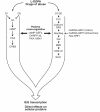
Simplified diagram of the protein kinase A (PKA) and extracellular signal-regulated kinase/mitogen-activated protein kinase (ERK1/2; MAPK) signaling cascades. Both PKA and ERK cascades control neuronal activity and immediate early gene (IEG) expression in medium spiny projection neurons of the striatum. D1 dopamine receptors promote the PKA cascade by activating adenylyl cyclase (AC) whereas D2 dopamine receptors inhibit AC. D1/D2 heterodimers are positively coupled to phospholipase C (PLC). The calcium- and diacylglycerol-regulated guanine nucleotide exchange factors (CalDAG-GEFs), which regulate the ERK1/2 cascade, are differentially expressed in the striosome and matrix compartments. Imbalances in striosome vs. matrix (IEG) transcription are implicated in l-DOPA-induced dyskinesias and drug addiction.
Figure 3
Figure 3
Models of striosome–matrix signaling imbalances in disease. Schematic diagram illustrating relative activity or cell density (dense stipple for high activity, sparse stipple for low activity) found in studies of human brain and brains of animal models. For the diseases or disease models listed, observations were made of either a relative imbalance in MSN cell density, immunomarker expression or IEG induction favoring the striosomes (A) or matrix (B), relative to the opposite compartment. The diagram is based on a cross-section through the striatum of an adult human, immunostained for choline acetyltransferase. Medial is to the right. CN, caudate nucleus; P, putamen; IC, internal capsule; LID, l-DOPA-induced dyskinesia; MSA-P, multiple system atrophy with parkinsonian features; HD, Huntington’s disease; DYT3, X-linked dystonia-parkinsonism; DRD, dopamine-responsive dystonia (DYT5).

References

    1. Adams D. H., Hanson G. R., Keefe K. A. (2001). Differential effects of cocaine and methamphetamine on neurotensin/neuromedin N and preprotachykinin messenger RNA expression in unique regions of the striatum. Neuroscience 102, 843–85110.1016/S0306-4522(00)00530-3 - DOI - PubMed
    1. Adams D. H., Hanson G. R., Keefe K. A. (2003). Distinct effects of methamphetamine and cocaine on preprodynorphin messenger RNA in rat striatal patch and matrix. J. Neurochem. 84, 87–9310.1046/j.1471-4159.2003.01507.x - DOI - PubMed
    1. Albin R. L., Young A. B., Penney J. B. (1989). The functional anatomy of basal ganglia disorders. Trends Neurosci. 12, 366–37510.1016/0166-2236(89)90074-X - DOI - PubMed
    1. Albin R. L., Young A. B., Penney J. B. (1995). The functional anatomy of disorders of the basal ganglia. Trends Neurosci. 18, 63–6410.1016/0166-2236(95)93943-R - DOI - PubMed
    1. Alexander G. E., Delong M. R., Strick P. L. (1986). Parallel organization of functionally segregated circuits linking basal ganglia and cortex. Annu. Rev. Neurosci. 9, 357–38110.1146/annurev.ne.09.030186.002041 - DOI - PubMed