. 2011 Jun;9(2):225-40.
doi: 10.2166/wh.2010.106.

Human norovirus infection of caco-2 cells grown as a three-dimensional tissue structure

Affiliations

Human norovirus infection of caco-2 cells grown as a three-dimensional tissue structure

Timothy M Straub et al. J Water Health. 2011 Jun.

Abstract

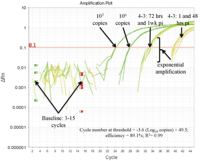
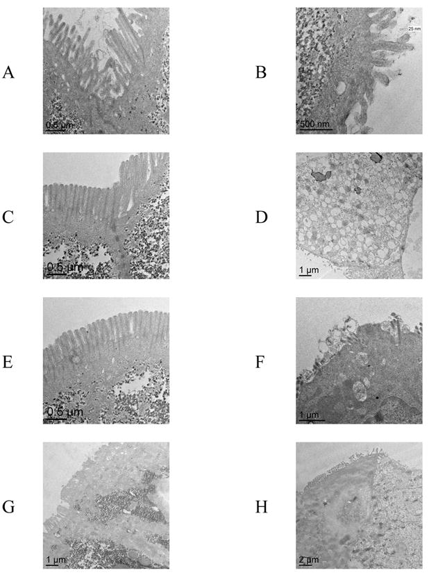
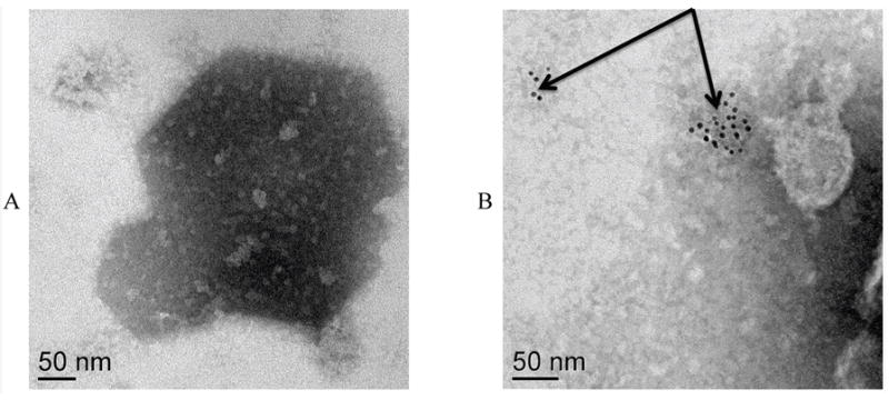
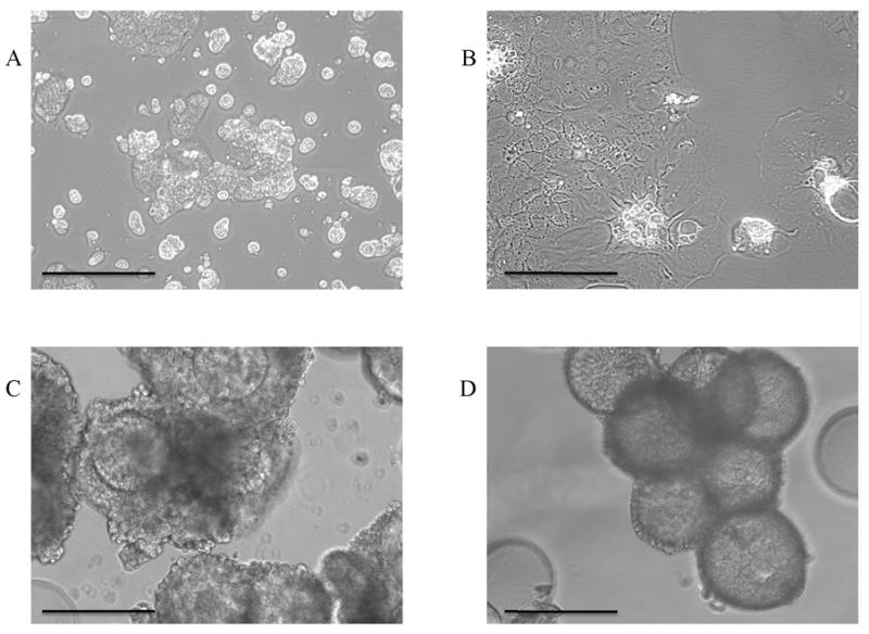
Human norovirus (hNoV) infectivity was studied using a three-dimensional model of large intestinal epithelium. Large intestine Caco-2 cells were grown in rotating wall vessel bioreactors for 18-21 days at 37 degrees C and then transferred to 24-well tissue culture plates where they were infected with GI.1 and GII.4 human noroviruses collected from human challenge trials and various outbreak settings, respectively. Compared with uninfected cells, transmission micrographs of norovirus-infected cells displayed evidence of shortening or total loss of apical microvilli, and vacuolization. Quantitative reverse transcription real-time PCR (qRT-PCR) indicated an approximate 2-3 log10 increase in viral RNA copies for the infected cells. A passage experiment examined both the ability for continued viral RNA and viral antigen detection. In the passaged samples 1.01x10(6) copies ml(-1) were detected by qRT-PCR. Immune electron microscopy using primary antibody to hNoV GI.1 capsids in conjunction with 6 nm gold-labelled secondary antibodies was performed on crude cellular lysates. Localization of antibody was observed in infected but not for uninfected cells. Our present findings, coupled with earlier work with the three-dimensional small intestinal INT407 model, demonstrate the utility of 3-D cell culture methods to develop infectivity assays for enteric viruses that do not readily infect mammalian cell cultures.


Figures

Figure 1
Figure 1
Logarithmic amplification plot of a quantitative reverse transcription real-time PCR experiment comparing hNoV infected Caco-2 cells with standards. For clarity only the 107 and 106 copy standards are displayed. The data for the infected cells is presented in Table 4. Note that the slopes of the samples vs. standards are similar in the exponential phase of amplification. This indicates that the samples had little to no PCR inhibition throughout the time course of the experiment. For all experiments, threshold for positive amplification was ΔRN = 0.1, and baseline was set between 3 and 15 PCR cycles.
Figure 2
Figure 2
Lack of gross pathology observed in NoV infected 3-D Caco-2 cells. Observations of the 3-D tissue aggregates under an inverted microscope showed little if any, indication of infection by hNoV. Samples 4-1 and 4-3 were stool samples from the same patient harvested pre (4-1) and post (4-3) infection with GI.1 hNoV from a previous human challenge trial. The left column of photos (A, C, E, G at 1, 24, 48, and 72 hrs post-challenge, respectively) show the course of challenge using 4-1), and the right column of photos (B, D, F, and H at1, 24, 48, and 72-hrs post-challenge, respectively) show the course of a challenge using 4-3. While (H, arrow) showed preliminary signs of infection, it was the exception rather than the rule and similar examples could be observed in uninfected controls at 72 hrs post challenge.
Figure 3
Figure 3
Transmission electron micrographs comparing stool sample challenges of the 3-D Caco-2 cells using 4-1 (negative) and 4-3 (hNoV positive) stool samples. The left column of photos (A, C, E, and G at1-hr, 24-hrs, 48 hrs, and 72-hrs post-challenge, respectively) show the course of a challenge using 4-1, and the right column of photos (B, D, F, and H at1-hr, 24-hrs, 48 hrs, and 72-hrs post-challenge, respectively) show the course of a challenge using 4-3. Note the presence of microvilli on all cells challenged with hNoV negative stool sample (left column of photos) and the presence of tight junctions (C). In (H), a relatively healthy cell with microvilli is adjacent to cells that are presumably infected with hNoV.
Figure 4
Figure 4
Immune electron microscopy of cell lysates from GI.1 hNoV infected 3-D Caco-2 cells (A) Failure of localization of secondary antibody when primary antibodies were omitted. (B) Localization of 6 nm gold secondary antibodies (arrows) when infectious samples received both primary and secondary antibody.
Figure 5
Figure 5
Observational phenotypes of Caco-2 cells grown in 75 cm2 flasks (A and B) and as dynamic 3 – dimensional cell cultures (C and D). Cells shown in A and C are from Washington State University, and cells shown in B and D were purchased directly from ATCC. Bar = 250 microns.

References

    1. Asanaka M, Atmar RL, Ruvolo V, Crawford SE, Neill FH, Estes MK. Replication and packaging of norwalk virus RNA in cultured cells. Proc Natl Acad Sci. 2005;102(29):10327–10332. - PMC - PubMed
    1. Atmar RL, Opeku AR, Gilger MA, Estes MK, Crawford SE, Neill FH, Graham DY. Norwalk virus shedding after experimental human infection. Emerg Infect Dis. 2008;14(10):1533–1537. - PMC - PubMed
    1. Carterson AJ, Honer zu Bentrup K, Ott CM, Clarke MS, Pierson DL, Vanderburg CR, Buchanan KL, Nickerson CA, Schurr MJ. A549 lung epithelial cells grown as three-dimensional aggregates: Alternative tissue culture model for Psuedomonas aeruginosa. Infect Immun. 2005;73(2):1129–1140. - PMC - PubMed
    1. Carvalho HM, Teel LD, Goping G, O’Brien AD. A three dimensional tissue culture model for the study of attach and efface lesion formation by enteropathogenic and enterohaemorrhagic Esherichia coli. Cell Microbiol. 2005;7:1771–1781. - PubMed
    1. Centers for Disease Control and Prevention (U.S.) Norovirus activity - United States, 2006-2007. MMWR Morb Mortal Wkly Rep. 2007;56(33):842–846. - PubMed

Publication types