Review
. 2012 Jun 15;16(12):1467-75.
doi: 10.1089/ars.2011.4277. Epub 2011 Dec 15.

Synaptic mitochondrial pathology in Alzheimer's disease

Affiliations
Review

Synaptic mitochondrial pathology in Alzheimer's disease

Heng Du et al. Antioxid Redox Signal. .

Abstract

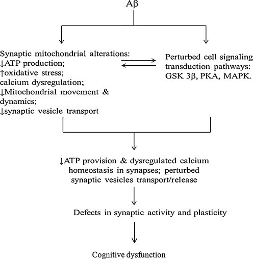
Significance: Synaptic degeneration, an early pathological feature in Alzheimer's disease (AD), is closely correlated to impaired cognitive function and memory loss. Recent studies suggest that involvement of amyloid-beta peptide (Aβ) in synaptic mitochondrial alteration underlies these synaptic lesions. Thus, to understand the Aβ-associated synaptic mitochondrial perturbations would fortify our understanding of synaptic stress in the pathogenesis of AD.

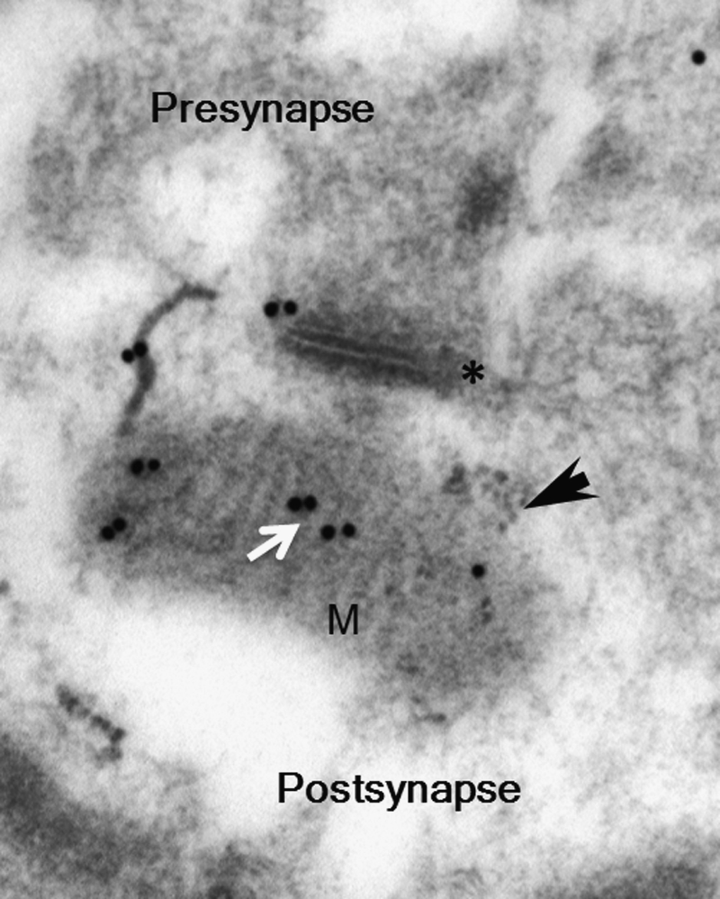
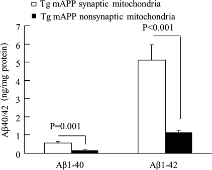
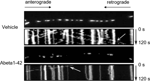
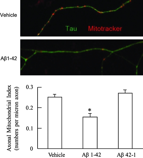
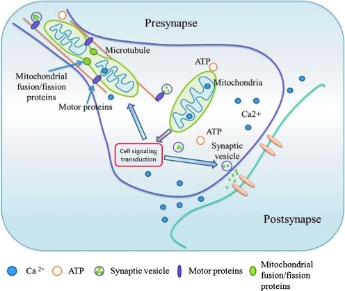
Recent advances: Increasing evidence suggests that synaptic mitochondrial dysfunction is strongly associated with synaptic failure in many neurodegenerative diseases including AD. Based on recent findings in human AD subjects, AD animal models, and AD cellular models, synaptic mitochondria undergo multiple malfunctions including Aβ accumulation, increased oxidative stress, decreased respiration, and compromised calcium handling capacity, all of which occur earlier than changes seen in nonsynaptic mitochondria before predominant AD pathology. Of note, the impact of Aβ on mitochondrial motility and dynamics exacerbates synaptic mitochondrial alterations.

Critical issues: Synaptic mitochondria demonstrate early deficits in AD; in combination with the role that synaptic mitochondria play in sustaining synaptic functions, deficits in synaptic mitochondria may be a key factor involved in an early synaptic pathology in AD.

Future directions: The importance of synaptic mitochondria in supporting synapses and the high vulnerability of synaptic mitochondria to Aβ make them a promising target of new therapeutic strategy for AD.


Figures

FIG. 1.
FIG. 1.
Accumulation of Aβ in synaptic mitochondria. Immunogold electron microscopy images with a specific Aβ1–42 antibody followed by gold-conjugated antibody (18 nm) to show the presence of intra-mitochondrial Aβ accumulation (white arrow) in 12-month-old Tg mAPP. The black arrow denotes mitochondria. The asterisk (*) denotes a synapse. M indicated mitochondria. Aβ, amyloid-beta peptide; APP, amyloid-beta precursor protein.
FIG. 2.
FIG. 2.
Synaptic mitochondria are more vulnerable to Aβ accumulation. Synaptic mitochondria and nonsynaptic mitochondria were prepared from Tg mAPP mice at age of 4 months and subjected to measure the levels of Aβ1–40 and Aβ1–42. The data showed that levels of both Aβ species were significantly increased in synaptic mitochondria.
FIG. 3.
FIG. 3.
Aβ affects axonal mitochondrial movement. Representative kymograph images of the vehicle and Aβ1–42-treated axonal mitochondrial movement showing injured axonal mitochondrial movement. The arrows denote the traces of mitochondrial movement.
FIG. 4.
FIG. 4.
Aβ alters axonal mitochondrial distribution. The axonal mitochondrial density is decreased after Aβ1–42 (200 nM) treatment for 24 h on cultured hippocampal neurons (Lower panel). *p<0.05 vs. cells treated with vehicle or reversed Aβ42-1. The upper panel shows representative images for vehicle- or Aβ-treated axonal mitochondrial distribution. Double immunostaining with Mitotracker (red, mitochondrial marker) and Tau (green, axonal marker) was performed. (To see this illustration in color the reader is referred to the web version of this article at www.liebertonline.com/ars).
FIG. 5.
FIG. 5.
Schematic figure to show the role of synaptic mitochondria in supporting synaptic activity. Synaptic mitochondria are generated in neuronal soma and further transported to synapses. Normal synaptic mitochondrial movement, docking, and dynamics are crucial features for mitochondria to exert their function on ATP production, calcium modulation, and regulation of cell signaling cascades, consequently maintaining synaptic plasticity and transmission. (To see this illustration in color the reader is referred to the web version of this article at www.liebertonline.com/ars).
FIG. 6.
FIG. 6.
Working hypothesis. In the presence of Aβ, mitochondrial transport and dynamics are injured with compromised synaptic mitochondrial structure and function, thus leading to decreased energy metabolism, dysregulated calcium homeostasis, and perturbed cell signaling cascades, eventually leading to synaptic injury and cognitive dysfunction.

References

    1. Alonso-Nanclares L. Gonzalez-Soriano J. Rodriguez JR. DeFelipe J. Gender differences in human cortical synaptic density. Proc Natl Acad Sci U S A. 2008;105:14615–14619. - PMC - PubMed
    1. Banaclocha MM. Hernandez AI. Martinez N. Ferrandiz ML. N-acetylcysteine protects against age-related increase in oxidized proteins in mouse synaptic mitochondria. Brain Res. 1997;762:256–258. - PubMed
    1. Billups B. Forsythe ID. Presynaptic mitochondrial calcium sequestration influences transmission at mammalian central synapses. J Neurosci. 2002;22:5840–5847. - PMC - PubMed
    1. Brown MR. Sullivan PG. Geddes JW. Synaptic mitochondria are more susceptible to Ca2+ overload than nonsynaptic mitochondria. J Biol Chem. 2006;281:11658–11668. - PubMed
    1. Buchanan ME. Davis RL. A distinct set of Drosophila brain neurons required for neurofibromatosis type 1-dependent learning and memory. J Neurosci. 2010;30:10135–10143. - PMC - PubMed

Publication types