Review
. 2012:7:123-44.
doi: 10.1146/annurev-pathol-011110-130227. Epub 2011 Sep 13.

Merkel cell carcinoma: a virus-induced human cancer

Affiliations
Review

Merkel cell carcinoma: a virus-induced human cancer

Yuan Chang et al. Annu Rev Pathol. 2012.

Abstract

Merkel cell polyomavirus (MCV) is the first polyomavirus directly linked to human cancer, and its recent discovery helps to explain many of the enigmatic features of Merkel cell carcinoma (MCC). MCV is clonally integrated into MCC tumor cells, which then require continued MCV oncoprotein expression to survive. The integrated viral genomes have a tumor-specific pattern of tumor antigen gene mutation that incapacitates viral DNA replication. This human cancer virus provides a new model in which a common, mostly harmless member of the human viral flora can initiate cancer if it acquires a precise set of mutations in a host with specific susceptibility factors, such as age and immune suppression. Identification of this tumor virus has led to new opportunities for early diagnosis and targeted treatment of MCC.


Figures

Figure 1
Figure 1
Clinical appearance of Merkel cell carcinoma (MCC) lesion. Red, raised, nodular MCC lesion on the arm of a patient. Reproduced courtesy of Dr. Klaus J. Busam, Department of Pathology, Memorial Sloan-Kettering Cancer Center, New York.
Figure 2
Figure 2
Phylogenetic analysis of large tumor antigens from 12 polyomaviruses. The nine known human polyomaviruses (red font) are BK virus (BKV: NC_001538); JC virus (JCV: NC_001699); Washington University polyomavirus (WUV: NC_009539), Karolinska Institute polyomavirus (KIV: NC_009238); Merkel cell polyomavirus (MCV: NC_010277); trichodysplasia spinulosum polyomavirus (TSaPyV: NC_014361); and human polyomaviruses (HPyV) 6 (NC_014406), 7 (NC_014407), and 9 (HQ696595). Also shown are several well-studied nonhuman polyomaviruses (gray font): simian vacuolating virus 40 (SV40: NC_001669), murine polyomavirus (MPyV: NC_001515), and African green monkey lymphotropic polyomavirus (LPV: NC_004763). The phylogenetic tree was produced using ClustalX (112, 113). Reproduced courtesy of Dr. Huichen Feng, Cancer Virology Program, Hillman Cancer Center, University of Pittsburgh Cancer Institute, Pittsburgh, Pennsylvania.
Figure 3
Figure 3
Genome organization of Merkel cell carcinoma. The wild-type Merkel cell polyomavirus genome is 5,387 bp long and contains a noncoding regulatory region (NCRR), from which the virus bidirectionally encodes for early and late proteins. In addition to the early and late transcriptional promoters, the NCRR also contains the viral origin (red), which can be mapped to 71 bp of contiguous sequence recognized by the large tumor antigen (LT). LT, small T antigen (sT), and 57-kT antigen constitute the gene products of the early region. Viral protein 1 (VP1) and VP2 are late proteins required for capsid formation and viral replication.
Figure 4
Figure 4
Genome, transcript, and protein features of the tumor antigen (T antigen) locus. Merkel cell polyomavirus (MCV) T antigens contain protein motifs similar to those found in other polyomaviruses (red font). In its amino-terminal half, preserved even in the presence of tumor-specific truncation mutations, the MCV large T antigen (LT) contains conserved region 1 (CR1)-, DnaJ-, and retinoblastoma (Rb)-binding amino acid sequences. In MCV, ~200 amino acids are inserted immediately following the first exon and before the Rb-binding site. This MCV-unique region (MUR) exhibits no significant homology to other polyomavirus T antigens, which suggests that it may confer unique functions. The C-terminal portion of MCV is occupied largely by motifs required for replication, including the origin-binding domain (OBD) and the zinc finger, leucine zipper, ATPase, and helicase domains. The small T antigen (sT) contains a protein phosphatase 2A (PP2A)-binding motif that is absent in LT and the 57-kT antigen because of the differential splicing of their respective transcripts.
Figure 5
Figure 5
Merkel cell carcinoma (MCC) pathology. (a) Monomorphous clusters of MCC tumor cells in dermis stained with hematoxylin and eosin (20× magnification). (b) Adjacent section immunostained with anti-CK20 antibody, demonstrating distinctive perinuclear localization in a dot-like pattern (20× magnification).
Figure 6
Figure 6
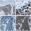
Expression of Merkel cell polyomavirus (MCV) tumor antigen (T antigen) proteins in Merkel cell carcinoma (MCC). (a) Immunostaining with anti-MCV large T antigen (LT) shows nests of MCC tumor cells infiltrating the dermis (20× magnification). (b) Oil emersion demonstrates the robust expression of MCV LT localized to the nuclear compartment of tumor cells (100× magnification). (c,d) Differential expression of LT and small T antigen (sT) in a case of MCC. Sequential sections of a polymerase chain reaction–confirmed, MCV DNA–positive MCC stained with (c) anti-MCV LT and (d) anti-MCV sT antibodies. Although LT expression was not detected in this lesion, sT expression is clearly seen. Slides are shown at 20× magnification with a hematoxylin counterstain.
Figure 7
Figure 7
Steps in the molecular evolution of Merkel cell carcinoma (MCC). (1) Merkel cell polyomavirus (MCV) infection is common in the general population and is acquired in early childhood. It is an easily detected component of the normal skin flora. (2) In the setting of decreased immune competence (either iatrogenically induced or age related), reactivation of MCV may occur, as has been observed with other human polyomaviruses. (3) Such a burst of infective virus production can facilitate viral integration in susceptible Merkel cells. (4) Selection pressure is predicted to occur against Merkel cells infected with replication-competent viruses as a result of cell lysis. (5) The expression of MCV tumor antigens (T antigens) in cells with integrated virus harboring T antigen–truncation mutations provides proproliferative signals that lead to MCC.
Figure 8
Figure 8
Merkel cell carcinoma (MCC) metastasis to a lymph node involved with chronic lymphocytic leukemia (CLL). Immunohistochemical study of a lymph node containing both MCC and CLL shows tumor antigen expression only in MCC tumor cells in the typical nuclear pattern, but not in CLL tumor cells. (a) 10× magnification. (b) 100× magnification, oil emersion, hematoxylin counterstain.

References

    1. Toker C. Trabecular carcinoma of the skin. Arch Dermatol. 1972;105:107–10. - PubMed
    1. Engels EA, Frisch M, Goedert JJ, Biggar RJ, Miller RW. Merkel cell carcinoma and HIV infection. Lancet. 2002;359:497–98. - PubMed
    1. Agelli M, Clegg LX. Epidemiology of primary Merkel cell carcinoma in the United States. J Am Acad Dermatol. 2003;49:832–41. - PubMed
    1. Hodgson NC. Merkel cell carcinoma: changing incidence trends. J Surg Oncol. 2005;89:1–4. - PubMed
    1. Feng H, Shuda M, Chang Y, Moore PS. Clonal integration of a polyomavirus in human Merkel cell carcinoma. Science. 2008;319:1096–100. - PMC - PubMed

Publication types

MeSH terms