. 2011 Sep 25:11:268.
doi: 10.1186/1471-2148-11-268.

A holistic phylogeny of the coronin gene family reveals an ancient origin of the tandem-coronin, defines a new subfamily, and predicts protein function

Affiliations

A holistic phylogeny of the coronin gene family reveals an ancient origin of the tandem-coronin, defines a new subfamily, and predicts protein function

Christian Eckert et al. BMC Evol Biol. .

Abstract

Background: Coronins belong to the superfamily of the eukaryotic-specific WD40-repeat proteins and play a role in several actin-dependent processes like cytokinesis, cell motility, phagocytosis, and vesicular trafficking. Two major types of coronins are known: First, the short coronins consisting of an N-terminal coronin domain, a unique region and a short coiled-coil region, and secondly the tandem coronins comprising two coronin domains.

Results: 723 coronin proteins from 358 species have been identified by analyzing the whole-genome assemblies of all available sequenced eukaryotes (March 2011). The organisms analyzed represent most eukaryotic kingdoms but also cover every taxon several times to provide a better statistical sampling. The phylogenetic tree of the coronin domains based on the Bayesian method is in accordance with the most recent grouping of the major kingdoms of the eukaryotes and also with the grouping of more recently separated branches. Based on this "holistic" approach the coronins group into four classes: class-1 (Type I) and class-2 (Type II) are metazoan/choanoflagellate specific classes, class-3 contains the tandem-coronins (Type III), and the new class-4 represents the coronins fused to villin (Type IV). Short coronins from non-metazoans are equally related to class-1 and class-2 coronins and thus remain unclassified.

Conclusions: The coronin class distribution suggests that the last common eukaryotic ancestor possessed a single and a tandem-coronin, and most probably a class-4 coronin of which homologs have been identified in Excavata and Opisthokonts although most of these species subsequently lost the class-4 homolog. The most ancient short coronin already contained the trimerization motif in the coiled-coil domain.


Figures

Figure 1
Figure 1
Phylogenetic tree of the coronin family. The phylogenetic tree of the coronin family was calculated from the multiple sequence alignment of the conserved coronin domain using the Bayesian method. The unrooted tree was drawn with iTOL [73] and branches were coloured according to class and taxonomic distributions. For an extended representation of the tree including all posterior probability values see Additional file 2.
Figure 2
Figure 2
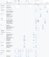
Coronin repertoire of selected species of major taxa and branches. The coronins of several representative species for most eukaryotic taxa and branches are listed (for the list of all species see Additional file 4). On top, alternatively used names and classification schemes are given for better comparison and orientation.
Figure 3
Figure 3
Domain organisation of representative coronins. A colour key to the domain names and symbols is given on the right except for the coronin domain that is coloured in orange. The abbreviations for the domains are: WD, WD repeat; PH, pleckstrin-homology domain; LZ, leucine zipper; VHP, villin headpeace domain.
Figure 4
Figure 4
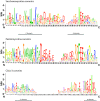
Sequence conservation in the CA domains. The sequence logos illustrate the sequence conservation within the multiple sequence alignments of the CA domains of the Saccharomycotina, the Pezizomycotina, and the class-3 coronins. The CA domains of the Saccharomycotina and the Pezizomycotina are located within the unique regions of the short coronins while the CA domain of the class-3 coronins is at the C-termini of the proteins like in WASP family proteins. The regions between the C and the A domains are of variable length.
Figure 5
Figure 5
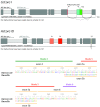
Gene structures of alternatively spliced coronins. The cartoons outline the gene structures of the alternatively spliced coronin-1 gene from Caenorhabditis elegans, CeCoro1, and the coronin-1D gene from Homo sapiens, HsCoro1D. The alternatively spliced CeCoro1 gene contains a differentially included exon8, which has an additional alternative 3'-splice site, leading to three transcripts. The other two described splice sites, an alternative 3'-splice site of exon7 and an alternative 5'-splice site of exon8 [35], are most probably artificial. The HsCoro1D gene contains a cluster of two mutually exclusive spliced exons, exon5a and exon5b, and an alternative 5'-splice site of exon10. Dark grey bars and light grey bars mark exons and introns, respectively, and alternative exons and splice sites are coloured.
Figure 6
Figure 6
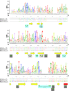
Sequence conservation within the actin binding region. The sequence logos illustrate the sequence conservation within the multiple sequence alignments of the coronin domains. Here, only the N- and C-termini of the coronin domains are shown because most of the residues implicated in actin binding map to these regions. For the representation of the entire coronin domain see Additional file 5. For better orientation, the sequences of three representative coronins are shown: the yeast coronin as the main target of mutagenesis experiments, the Dictyostelium coronin as the founding member of the protein family, and the murine coronin-1A of which the crystal structure is known. Secondary structural elements as determined from the crystal structure are drawn as yellow arrows (β-strands) and red boxes (α-helices). Green dots point to amino acids of ScCoro that have been mutated to alanine [41] and red dots highlight mutagenesis studies in HsCoro1B [40]. Light-blue boxes highlight mutations that abolished actin binding, dark-grey boxes represent mutations that did not influence actin binding, and light-grey boxes point to mutations in yeast coronins that could not be expressed and tested.
Figure 7
Figure 7
Evolution of the coronin protein family with respect to the species evolution. Schematic representation of the most widely accepted eukaryotic tree of life. Branch lengths are arbitrary. The coronin inventories of certain taxa and specific species have been plotted to the tree with class numbers given in colour-coded boxes. "O" stands for "Orphan", the unclassified short coronins. The numbers on the arrows refer to alternative placing of the respective taxa: 1: The independence of the Diplomonadida (instead of grouping them to the superkingdom Excavata) is supported by [51]. 2: The monophyly of the Rhodophyta is supported by [28,55]. 3: Grouping the Haptophyceae and Cryptophyta to the SAR is supported by [55-57].
Figure 8
Figure 8
Evolution of the coronin classes. The cartoon shows the different gene duplication and fusion events that led to the formation of the short coronins, the class-3 coronins, and the class-4 coronins.

References

    1. de Hostos EL, Bradtke B, Lottspeich F, Guggenheim R, Gerisch G. Coronin, an actin binding protein of Dictyostelium discoideum localized to cell surface projections, has sequence similarities to G protein beta subunits. EMBO J. 1991;10:4097–4104. - PMC - PubMed
    1. Tardieux I, Liu X, Poupel O, Parzy D, Dehoux P, Langsley G. A Plasmodium falciparum novel gene encoding a coronin-like protein which associates with actin filaments. FEBS Lett. 1998;441:251–256. doi: 10.1016/S0014-5793(98)01557-9. - DOI - PubMed
    1. Figueroa JV, Precigout E, Carcy B, Gorenflot A. Identification of a coronin-like protein in Babesia species. Ann N Y Acad Sci. 2004;1026:125–138. doi: 10.1196/annals.1307.017. - DOI - PubMed
    1. Heil-Chapdelaine RA, Tran NK, Cooper JA. The role of Saccharomyces cerevisiae coronin in the actin and microtubule cytoskeletons. Curr Biol. 1998;8:1281–1284. doi: 10.1016/S0960-9822(07)00539-8. - DOI - PubMed
    1. Suzuki K, Nishihata J, Arai Y, Honma N, Yamamoto K, Irimura T, Toyoshima S. Molecular cloning of a novel actin-binding protein, p57, with a WD repeat and a leucine zipper motif. FEBS Lett. 1995;364:283–288. doi: 10.1016/0014-5793(95)00393-N. - DOI - PubMed

Publication types

LinkOut - more resources