

Diagnosis of pericardial cysts using diffusion weighted magnetic resonance imaging: A case series
- PMID: 21943086
- PMCID: PMC3189152
- DOI: 10.1186/1752-1947-5-479
Diagnosis of pericardial cysts using diffusion weighted magnetic resonance imaging: A case series
Abstract
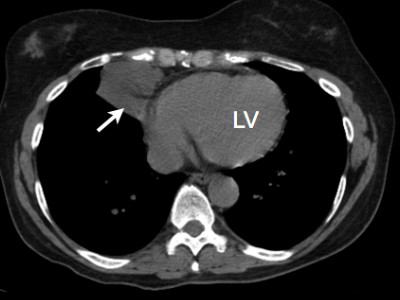
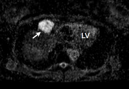
Introduction: Congenital pericardial cysts are benign lesions that arise from the pericardium during embryonic development. The diagnosis is based on typical imaging features, but atypical locations and signal magnetic resonance imaging sequences make it difficult to exclude other lesions. Diffusion-weighted magnetic resonance imaging is a novel method that can be used to differentiate tissues based on their restriction to proton diffusion. Its use in differentiating pericardial cysts from other pericardial lesions has not yet been described.
Case presentation: We present three cases (a 51-year-old Caucasian woman, a 66-year-old Caucasian woman and a 77-year-old Caucasian woman) with pericardial cysts evaluated with diffusion-weighted imaging using cardiac magnetic resonance imaging. Each lesion demonstrated a high apparent diffusion coefficient similar to that of free water.
Conclusion: This case series is the first attempt to investigate the utility of diffusion-weighted magnetic resonance imaging in the assessment of pericardial cysts. Diffusion-weighted imaging may be a useful noninvasive diagnostic tool for pericardial cysts when conventional imaging findings are inconclusive.
Figures
References
-
- McAllister HA Jr. Primary tumors and cysts of the heart and pericardium. Curr Probl Cardiol. 1979;4:1–51. - PubMed
LinkOut - more resources
Full Text Sources
