. 2012 Mar;165(6):1891-1903.
doi: 10.1111/j.1476-5381.2011.01677.x.

A novel angiopoietin-derived peptide displays anti-angiogenic activity and inhibits tumour-induced and retinal neovascularization

Affiliations

A novel angiopoietin-derived peptide displays anti-angiogenic activity and inhibits tumour-induced and retinal neovascularization

G M Palmer et al. Br J Pharmacol. 2012 Mar.

Abstract

Background and purpose: Pathological angiogenesis is associated with various human diseases, such as cancer, autoimmune diseases and retinopathy. The angiopoietin (Ang)-Tie2 system plays critical roles in several steps of angiogenic remodelling. Here, we have investigated the anti-angiogenic effect of a novel angiopoietin-derived peptide.

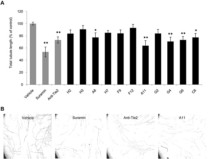
Experimental approach: Using computational methods, we identified peptides from helical segments within angiopoietins, which were predicted to inhibit their activity. These peptides were tested using biochemical methods and models of angiogenesis. The peptide with best efficacy, A11, was selected for further characterization as an anti-angiogenic compound.

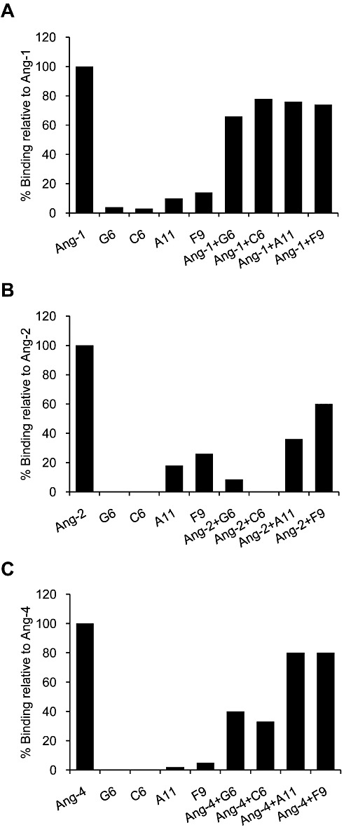
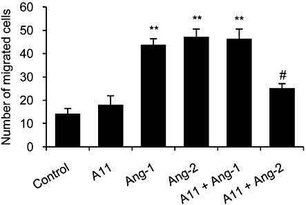
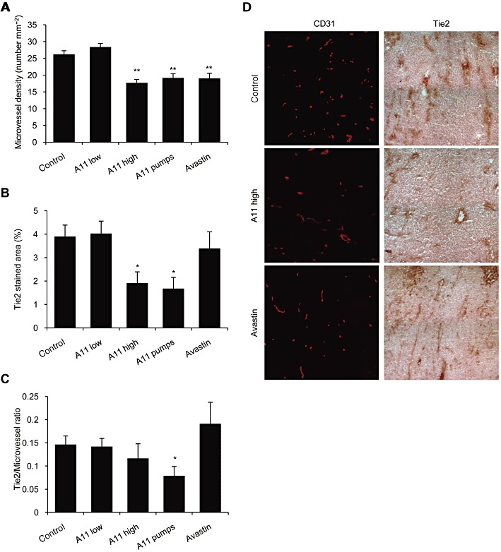
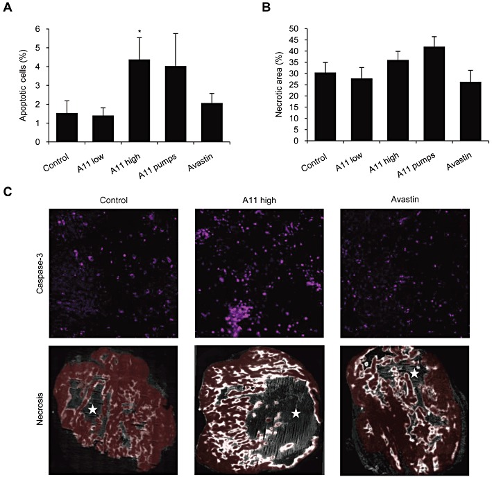
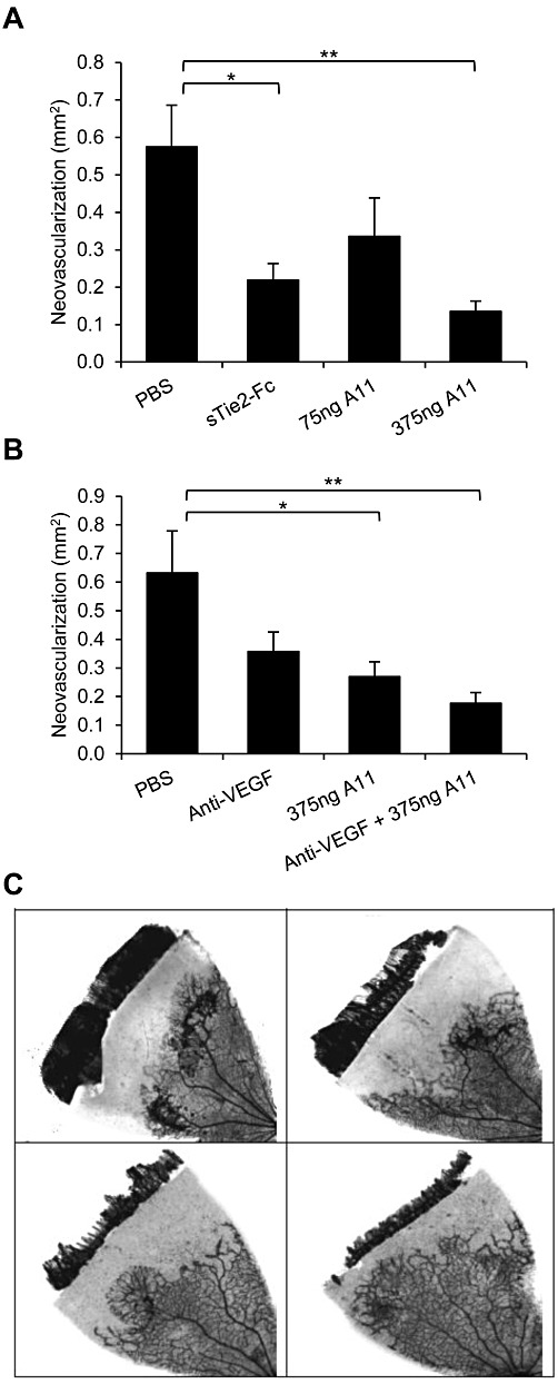
Key results: The potent anti-angiogenic activity of A11 was demonstrated in a multicellular assay of angiogenesis and in the chorioallantoic membrane model. A11 bound to angiopoietins and reduced the binding of Ang-2 to Tie2. A11 was also significantly reduced vascular density in a model of tumour-induced angiogenesis. Its ability to inhibit Ang-2 but not Ang-1-induced endothelial cell migration, and to down-regulate Tie2 levels in tumour microvessels, suggests that A11 targets the Ang-Tie2 pathway. In a rat model of oxygen-induced retinopathy, A11 strongly inhibited retinal angiogenesis. Moreover, combination of A11 with an anti-VEGF antibody showed a trend for further inhibition of angiogenesis, suggesting an additive effect.

Conclusions and implications: Our results indicate that A11 is a potent anti-angiogenic compound, through modulation of the Ang-Tie2 system, underlining its potential as a therapeutic agent for the treatment of ocular and tumour neovascularization, as well as other pathological conditions that are dependent on angiogenesis.


Figures

Figure 1
Figure 1
Peptides H2, H3, A8, H7, G4, G6, F9, F12, C6, A11 and G2 inhibit in vitro tubule formation. Co-cultures of human endothelial and interstitial cells (Angiokit) were treated in duplicates with 20 µM suramin, 5 µg·mL−1 anti-Tie2 neutralizing antibody or 1 µg·mL−1 of peptides. On day 14, cultures were fixed and stained for CD31, and tubule length was quantified. (A) Results are displayed as mean ± SEM of total tubule length relative to vehicle-treated cells (defined as 100%) and reflect the average obtained from four images in duplicate (total of eight measurements). *P < 0.05, **P < 0.01 significantly different from vehicle control; one-way ANOVA followed by Dunnett's post hoc test. (B) Representative images of cultures treated with vehicle control, suramin, anti-Tie2 mAb and peptide A11.
Figure 2
Figure 2
Inhibition of angiopoietin binding to Tie2. SPR analysis of angiopoietins binding to immobilized Tie2, in the presence or absence of peptides. The angiopoietin ligands and the four peptides tested, G6, C6, A11 and F9, were pre-incubated alone or together, and their ability to bind to immobilized Tie2 was measured. Results are shown for Ang-1 (A), Ang-2 (B) and Ang-4 (C) and are expressed relative to the binding to Tie2 of the respective ligand alone (defined as 100%).
Figure 3
Figure 3
Inhibition of in ovo angiogenesis in the CAM model. Tested compounds were applied on a restricted area of the CAM of fertilized eggs (n = 17–25). Peptides were tested at 0.5 and 5 nmol per egg. Fumagillin and anti-Tie2 neutralizing antibody were used as positive controls, at 5 and 0.4 µg per egg respectively. Forty-eight hours after treatment, CAMs were fixed, and total length of vessels was measured with image analysis software. Data are presented as mean ± SEM and expressed as % of vehicle control. **P < 0.01 significantly different from vehicle control; one-way ANOVA followed by Dunnett's post hoc test.
Figure 4
Figure 4
Inhibition of endothelial cell migration. HUVECs were serum-starved for 5 h, after which migration was induced by 250 ng·mL−1 of Ang-1 or Ang-2 in the presence or absence of A11. Cells were pre-treated with A11 (5 µg·mL−1) for 30 min. HUVECs were allowed to migrate for 4 h at 37°C, after which migrated cells were fixed, stained and scored. Data are presented as mean ± SEM and expressed as % of vehicle control. **P < 0.01 significantly different from vehicle control; one-way ANOVA followed by Dunnett's post hoc test. #P < 0.01 significantly different from Ang-2 alone; one-way ANOVA followed by Bonferroni's post hoc test.
Figure 5
Figure 5
Effects of systemic treatment with A11 on tumour angiogenesis and Tie2 levels. HCT116 human colorectal cancer cells were injected to the flank of nude mice. Animals were treated with A11 using twice daily i.p. injections of 100 µg (high) or 33 µg (low) or by continuous infusion (pumps) of 48 µg per day for 14 days. Bevacizumab (Avastin) given i.p. every other day for 14 days was used as a positive control. Frozen tumours were cryosectioned and stained. (A) Tumour microvessels were visualized by staining with anti-CD31 antibodies. Microvessel density is presented as means ± SEM number of vessels per area. **P < 0.01 significantly different from vehicle control; one-way ANOVA followed by Dunnett's post hoc test. (B) Tie2 protein levels were evaluated using anti-Tie2 antibodies and are presented as mean ± SEM percent of stained area. *P < 0.05 significantly different from vehicle control; one-way ANOVA followed by Dunnett's post hoc test. (C) The ratio between Tie2 levels and microvessel number is presented as means ± SEM. *P < 0.05 significantly different from vehicle control; Student's t-test. (D) Representative images of immunohistochemical staining of angiogenesis (anti-CD31) and Tie2 levels (anti-Tie2).
Figure 6
Figure 6
Effects of systemic treatment with A11 on tumour apoptosis and necrosis. HCT116 human colorectal cancer cells were injected to the flank of nude mice. Animals were treated with A11 using twice daily i.p. injections of 100 µg (high) or 33 µg (low) or by continuous infusion (pumps) of 48 µg per day for 14 days. Bevacizumab (Avastin) given i.p. every other day for 14 days was used as a positive control. Frozen tumours were cryosectioned and stained. (A) Apoptosis was followed using anti-cleaved caspase-3 antibodies and is presented as mean ± SEM percent of stained area. *P < 0.05 significantly different from vehicle control (t-test). (B) Necrosis was evaluated using EF5 immunofluorescent image (unstained necrotic regions were surrounded by brightly stained hypoxic regions). Results are presented as mean ± SEM percent of necrotic area within the analysed section (viable tumour plus necrotic area). (C) Representative images of immunohistochemical staining of apoptosis (cleaved caspase-3) and necrosis (EF5). Red colour marks the viable tumour tissue. Stars mark the necrotic areas.
Figure 7
Figure 7
Effect of peptide A11 on retinal neovascular growth. Retinal neovascularization was induced and assessed in Sprague–Dawley rats, using the OIR model. Data represent areas of abnormal vascular growth and are presented as mean ± SEM. (A) Rats received intravitreal injections of vehicle (PBS), peptide A11 at 75 or 375 ng per eye, or sTie2/Fc at 500 ng per eye as positive control. Both A11 at the higher dose and sTie-2/Fc yielded inhibition relative to PBS-injected eyes; **P < 0.01, *P < 0.05, significantly different as indicated; one-way ANOVA followed by Dunnett's post hoc test. (B). Rats received intravitreal injections of vehicle (PBS), anti-VEGF 500 ng per eye, peptide A11 at 375 ng per eye, or a combination of anti-VEGF and peptide A11 at 500 ng per eye and 375 ng per eye, respectively. Both peptide A11 and the combination of peptide A11 and anti-VEGF yielded inhibition relative to PBS-injected eyes; *P < 0.05, **P < 0.01, significantly different as indicated; one-way ANOVA followed by Dunnett's post hoc test. (C) Representative images of the degree of retinal neovascularization in the OIR model, following intravitreal injection of PBS (upper left), anti-VEGF (upper right), A11 (lower left) and A11 plus anti-VEGF (lower right).

References

    1. Asahara T, Chen D, Takahashi T, Fujikawa K, Kearney M, Magner M, et al. Tie2 receptor ligands, angiopoietin-1 and angiopoietin-2, modulate VEGF-induced postnatal neovascularization. Circ Res. 1998;83:233–240. - PubMed
    1. Augustin HG, Koh GY, Thurston G, Alitalo K. Control of vascular morphogenesis and homeostasis through the angiopoietin-Tie system. Nat Rev Mol Cell Biol. 2009;10:165–177. - PubMed
    1. Barnett JM, Yanni SE, Penn JS. The development of the rat model of retinopathy of prematurity. Doc Ophthalmol. 2010;120:3–12. - PMC - PubMed
    1. Bishop ET, Bell GT, Bloor S, Broom IJ, Hendry NF, Wheatley DN. An in vitro model of angiogenesis: basic features. Angiogenesis. 1999;3:335–344. - PubMed
    1. Brindle NP, Saharinen P, Alitalo K. Signaling and functions of angiopoietin-1 in vascular protection. Circ Res. 2006;98:1014–1023. - PMC - PubMed

MeSH terms