. 2011 Sep 23:1:30.
doi: 10.1186/2044-5040-1-30.

ColVI myopathies: where do we stand, where do we go?

Affiliations

ColVI myopathies: where do we stand, where do we go?

Valérie Allamand et al. Skelet Muscle. .

Abstract

Collagen VI myopathies, caused by mutations in the genes encoding collagen type VI (ColVI), represent a clinical continuum with Ullrich congenital muscular dystrophy (UCMD) and Bethlem myopathy (BM) at each end of the spectrum, and less well-defined intermediate phenotypes in between. ColVI myopathies also share common features with other disorders associated with prominent muscle contractures, making differential diagnosis difficult. This group of disorders, under-recognized for a long time, has aroused much interest over the past decade, with important advances made in understanding its molecular pathogenesis. Indeed, numerous mutations have now been reported in the COL6A1, COL6A2 and COL6A3 genes, a large proportion of which are de novo and exert dominant-negative effects. Genotype-phenotype correlations have also started to emerge, which reflect the various pathogenic mechanisms at play in these disorders: dominant de novo exon splicing that enables the synthesis and secretion of mutant tetramers and homozygous nonsense mutations that lead to premature termination of translation and complete loss of function are associated with early-onset, severe phenotypes. In this review, we present the current state of diagnosis and research in the field of ColVI myopathies. The past decade has provided significant advances, with the identification of altered cellular functions in animal models of ColVI myopathies and in patient samples. In particular, mitochondrial dysfunction and a defect in the autophagic clearance system of skeletal muscle have recently been reported, thereby opening potential therapeutic avenues.


Figures
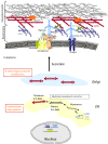
Figure 1
Figure 1
Schematic representation of the collagen type VI (ColVI) intracellular assembly process, and interactions with skeletal muscle extracellular matrix (ECM) components. Individual α(VI) chains fold through their triple helical domains to form monomers (1:1:1 ratio) in the endoplasmic reticulum (ER), which further align in an anti-parallel manner as dimers and tetramers that are stabilized by disulfide bonds between cysteine residues (S = S links). Post-translational modifications (indicated in orange) take place in the ER and Golgi, followed by secretion of tetramers that align non-covalently end to end, to form beaded microfibrils in the ECM. ColVI interacts with collagenous and non-collagenous components of the basal lamina and interstitial matrix surrounding muscle fibers.
Figure 2
Figure 2
Clinical spectrum, associated spine deformation and muscle MRI in collagen type VI (ColVI) myopathies. (A), Early severe phenotypes, corresponding to classic Ullrich congenital muscular dystrophy (UCMD), (B) intermediate forms seen in children or adults and (C) less severe, classic Bethlem myopathy (BM) forms constitute the overlapping clinical presentations of ColVI myopathies. (D) Radiography showing the evolution of spine deformation in a patient presenting with a classic early-onset UCMD phenotype. T1 transverse section of Bethlem myopathy upper limb girdle (E) and (F) thighs. Note the fatty infiltration, which appears as hyperintense area on T1-weighted images, located around the triceps brachialis muscles in (E) and along the fascia of vastus lateralis and vastus medialis muscles in (F) (yellow arrows). (G) The concentric fatty involvement of the thigh muscles is also seen on whole body MRI. (Images courtesy of Drs Susana Quijano-Roy and Tanya Stojkovic).
Figure 3
Figure 3
Collagen type VI (ColVI) expression study in cultured skin fibroblasts. (A) Representative images obtained using the protocol from [67] in which ColVI (red) is labeled with monoclonal antibody MAB1944 (Chemicon (now Millipore), Billerica, MA, USA) and perlecan (green) with monoclonal antibody MAB1948 (Chemicon). Note that ColVI expression appeared clearly altered in a patient with an early severe (ES) form and less so in patients with intermediate (Int) or Bethlem myopathy (BM) forms, compared with control fibroblasts (Cont). (B) Using the protocol of Hicks et al. [64], which detects ColVI (red) with polyclonal antibody Ab6588 (Abcam, Cambridge, UK) and fibronectin (green) with monoclonal antibody F15 (Sigma Chemical Co., St Louis, MO, USA), the sensitivity of the method is increased, and defective ColVI secretion could be detected in all patients' samples. Insets indicate nuclei, labeled using DAPI. Bars are 50 μm. (Images courtesy of Corine Gartioux and Valérie Allamand).
Figure 4
Figure 4
Repartition of the various types of mutations identified in the COL6A1, COL6A2 and COL6A3 genes. This schematic reflects, to the best of our ability, the distribution of 258 allelic mutations (98 on COL6A1, 113 on COL6A2 and 47 on COL6A3). Dominant de novo mutations represent 67% of the missense mutations, affecting glycine residues in the TH domains (Gly in TH), 57% of small deletions (< 5 amino acids) and 44% of splice-site mutations leading to in-frame exon skipping, whereas 97% of mutations leading to premature termination codons (PTCs) are familial (recessive or dominant).
Figure 5
Figure 5
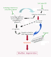
Current pathological hypotheses and therapeutic targets. The currently known cascade of main events leading to myofiber degeneration in ColVI-deficient skeletal muscle is shown. Mitochondrial dysfunction (due in part to the defective permeability transition pore (PTP) opening) triggers an energetic imbalance with the increased levels of phosphorylated adenosine monophosphate-activated protein kinase (p-AMPK), Ca2+ overload and the production of reactive oxygen species (ROS). Lack of autophagy induction exacerbates the cellular dysfunction because defective mitochondria and proteins (such as p62 aggregates) are not cleared from the cytoplasm. Together, these defects lead to increased apoptosis. Potential therapeutic interventions are indicated in green.

References

    1. Leitinger B, Hohenester E. Mammalian collagen receptors. Matrix Biol. 2007;26:146–155. doi: 10.1016/j.matbio.2006.10.007. - DOI - PubMed
    1. Bidanset D, Guidry C, Rosenberg L, Choi H, Timpl R, Hook M. Binding of the proteoglycan decorin to collagen type VI. J Biol Chem. 1992;267:5250–5256. - PubMed
    1. Bonaldo P, Russo V, Bucciotti F, Doliana R, Colombatti A. Structural and functional features of the alpha 3 chain indicate a bridging role for chicken collagen VI in connective tissues. Biochemistry. 1990;29:1245–1254. doi: 10.1021/bi00457a021. - DOI - PubMed
    1. Burg MA, Tillet E, Timpl R, Stallcup WB. Binding of the NG2 proteoglycan to type VI collagen and other extracellular matrix molecules. J Biol Chem. 1996;271:26110–26116. doi: 10.1074/jbc.271.42.26110. - DOI - PubMed
    1. Doane KJ, Yang G, Birk DE. Corneal cell-matrix interactions: type VI collagen promotes adhesion and spreading of corneal fibroblasts. Exp Cell Res. 1992;200:490–499. doi: 10.1016/0014-4827(92)90200-R. - DOI - PubMed