. 2011 Sep 21;101(6):1326-34.
doi: 10.1016/j.bpj.2011.08.004. Epub 2011 Sep 20.

Multiscale computational models for optogenetic control of cardiac function

Affiliations

Multiscale computational models for optogenetic control of cardiac function

Oscar J Abilez et al. Biophys J. .

Abstract

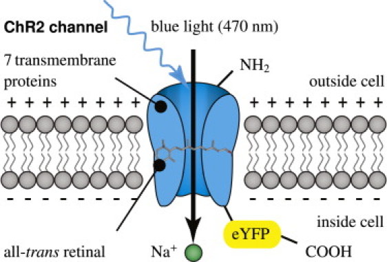
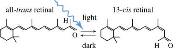
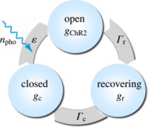
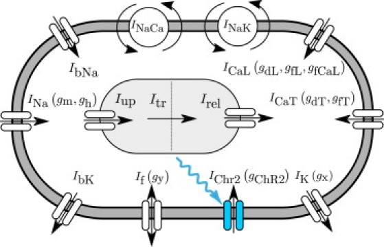
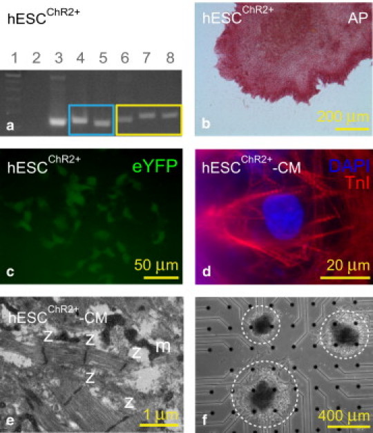
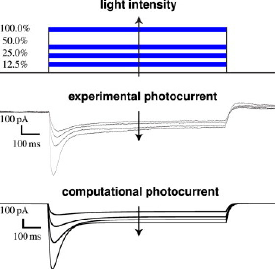
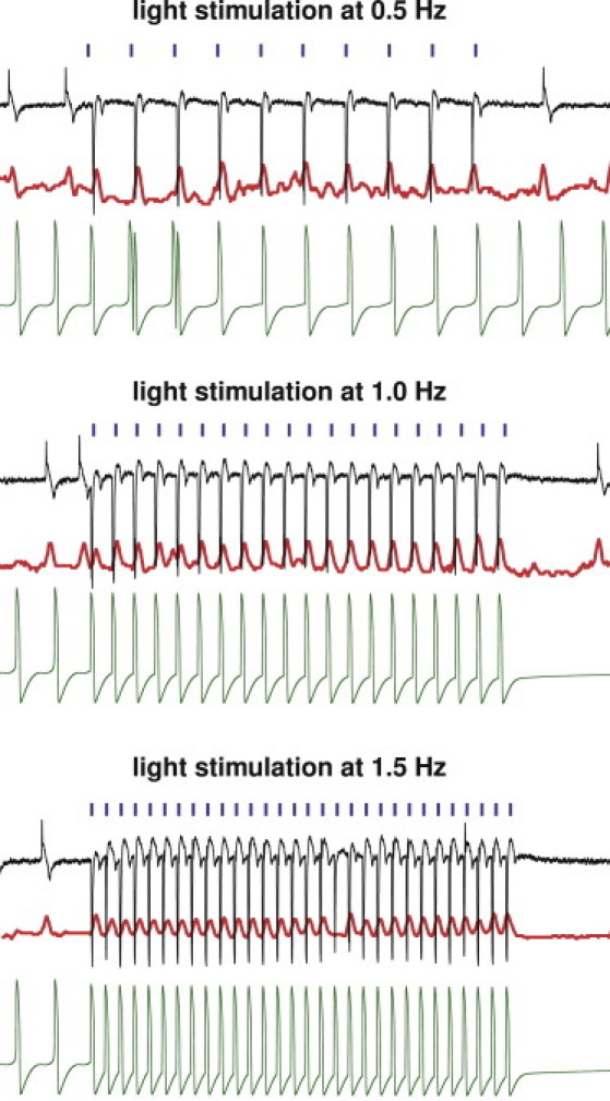
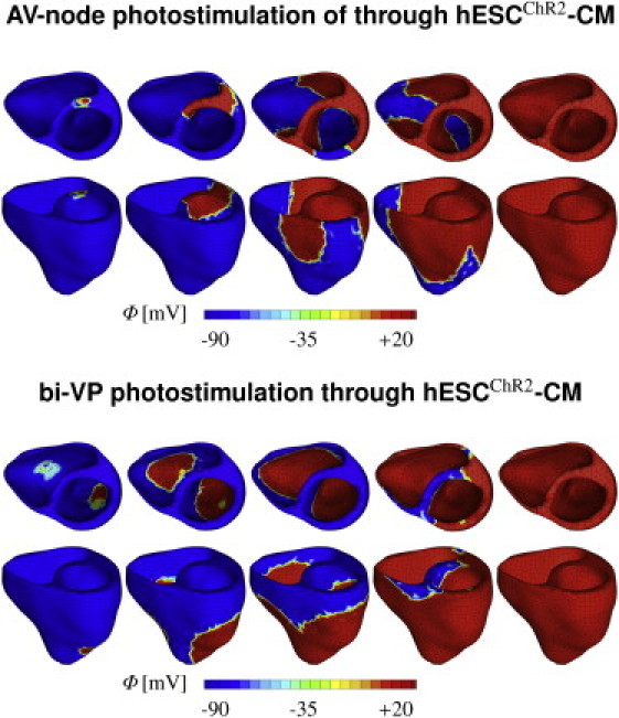
The ability to stimulate mammalian cells with light has significantly changed our understanding of electrically excitable tissues in health and disease, paving the way toward various novel therapeutic applications. Here, we demonstrate the potential of optogenetic control in cardiac cells using a hybrid experimental/computational technique. Experimentally, we introduced channelrhodopsin-2 into undifferentiated human embryonic stem cells via a lentiviral vector, and sorted and expanded the genetically engineered cells. Via directed differentiation, we created channelrhodopsin-expressing cardiomyocytes, which we subjected to optical stimulation. To quantify the impact of photostimulation, we assessed electrical, biochemical, and mechanical signals using patch-clamping, multielectrode array recordings, and video microscopy. Computationally, we introduced channelrhodopsin-2 into a classic autorhythmic cardiac cell model via an additional photocurrent governed by a light-sensitive gating variable. Upon optical stimulation, the channel opens and allows sodium ions to enter the cell, inducing a fast upstroke of the transmembrane potential. We calibrated the channelrhodopsin-expressing cell model using single action potential readings for different photostimulation amplitudes, pulse widths, and frequencies. To illustrate the potential of the proposed approach, we virtually injected channelrhodopsin-expressing cells into different locations of a human heart, and explored its activation sequences upon optical stimulation. Our experimentally calibrated computational toolbox allows us to virtually probe landscapes of process parameters, and identify optimal photostimulation sequences toward pacing hearts with light.


Figures

Figure 1
Figure 1
Channelrhodopsin-2 (ChR2) is a light-gated cation channel native to the green alga C. reinhardtii. It consists of seven transmembrane proteins and absorbs light through its interaction with retinal. Here, we induce channelrhodopsin coupled to enhanced yellow fluorescent protein (eYFP) into undifferentiated human embryonic stem cells via a lentiviral vector and differentiate these cells into cardiomyocytes.
Figure 2
Figure 2
Channelrhodopsin-2 (ChR2) is activated by photoisomerization of all-trans retinal to 13-cis retinal at wavelengths of 470 nm. After photoisomerization, the covalently bound retinal spontaneously relaxes to all-trans in the dark, providing closure of the ion channel and regeneration of the chromophore.
Figure 3
Figure 3
Three-state model for the channelrhodopsin photocycle. Upon photoabsorption, molecules in the closed state gc undergo a fast transition into the open state gChR2. After for some time, molecules spontaneously turn into the recovering state gr where the ion channels are closed, but the molecules are not yet ready to photoswitch again. After a recovery period, the molecules finally return to the closed state gc, ready to undergo a new photocycle when subjected to light.
Figure 4
Figure 4
Ionic model of genetically engineered light sensitive cardiac cell. The electrochemical state of the cell is characterized in terms of nion = 8 ion concentrations, cion = [ceNa, ceK, ceCa, ciNa, ciK, ciCa, cupCa, crelCa], the extracellular and intracellular sodium, potassium, and calcium concentrations, and the sarcoplasmic reticulum calcium uptake and release. Ion concentrations are controlled through ncrt = 12 ionic currents, Icrt = [INa, IbNa, If, INaCa, INaK, IChR2, IK, IbK, ICaL, ICaT, Iup, Irel], where the baseline autorhythmic cell model (26) has been enhanced with the channelrhodopsin current IChR2 (shown in blue). The channels are governed by ngate = 10 gating variables ggate = [gm, gh, gChR2, gy, gx, gdL, gfL, gfCa, gdT, gfT], which may be functions of the current membrane potential ϕ.
Figure 5
Figure 5
Undifferentiated human embryonic stem cells (hESC) stably transduced with a ChR2-eYFP lentiviral vector (hESCChR2+) remain pluripotent and can differentiate into cardiomyocytes (hESCChR2+-CM). (a) PCR shows that hESC ChR2+ express the pluripotent Oct-4 gene (169 bp, lane 4) and Nanog gene (154 bp, lane 5) (blue box). In addition, amplification within the ChR2 gene (174 bp, lane 6), across the ChR2-eYFP gene (197 bp, lane 7), and within the eYFP gene (187 bp, lane 8), confirms stable transduction of the ChR2-eYFP lentivirus in undifferentiated hESCChR2+ (yellow box). A ladder (100 bp, lane 1) confirms the predicted sizes of PCR products. Nontemplate control (lane 2) and GAPDH (152 bp, lane 3) serve as negative and positive controls, respectively. (b) Pluripotent hESCChR2+ stain is positive for alkaline phosphatase (red). (c) Fluorescence microscopy shows hESCChR2+ has a positive eYFP signal (green). (d) hESCChR2+-CM have positive TnI signals (red), consistent with a CM phenotype. DAPI staining (blue) demonstrates the position of nuclei. (e) Transmission electron microscopy shows sarcomeres with associated z-lines (z) and mitochondria (m) in hESCChR2+-CM. (f) Light microscopy shows three hESCChR2+-CM colonies (dashed white circles) on a multielectrode array.
Figure 6
Figure 6
Experimental and computational sensitivity of hESCChR2-CM with respect to light intensity. With light on, the photocurrent IChR2 increases rapidly, peaks, and decays toward a characteristic plateau value. With light off, the photocurrent IChR2 drops rapidly and decays to zero. Light intensity is varied from 12.5% to 25%, 50%, and 100% (top). Whole-cell voltage-clamp reveals an increased photocurrent IChR2 as the light intensity increases (middle). The computational hESCChR2-CM model captures the light sensitivity and displays increased photocurrents IChR2 with increased light intensity (bottom).
Figure 7
Figure 7
Experimental and computational sensitivity of hESCChR2-CM with respect to stimulation frequency. Light stimulation (blue) evokes field potentials (black) that translate into mechanical contractions (red). Light stimulation at 100% intensity is performed at 0.5 Hz (top), 1.0 Hz (middle), and 1.5 Hz (bottom). Evoked signals during light stimulation (center) are markedly different from pre- and poststimulation signals at all frequencies (left and right). The computational hESCChR2-CM model (green) captures the electrical signal at all frequencies, both during light stimulation (center), and pre- and poststimulation (left and right).
Figure 8
Figure 8
Virtual activation sequences of light-paced hearts. Atrioventricular node (top) and biventricular (bottom) photosimulations are initiated through hESCChR2-CM, virtually injected into the basal septum and into both lateral walls, respectively. All other regions are modeled as standard ventricular CM. The color code indicates the magnitude of the transmembrane potential ϕ varying from −90 mV (blue) to +20 mV (red).

References

    1. Oesterhelt D., Stoeckenius W. Rhodopsin-like protein from the purple membrane of Halobacterium halobium. Nat. New Biol. 1971;233:149–152. - PubMed
    1. Matsuno-Yagi A., Mukohata Y. Two possible roles of bacteriorhodopsin; a comparative study of strains of Halobacterium halobium differing in pigmentation. Biochem. Biophys. Res. Commun. 1977;78:237–243. - PubMed
    1. Nagel G., Ollig D., Hegemann P. Channelrhodopsin-1: a light-gated proton channel in green algae. Science. 2002;296:2395–2398. - PubMed
    1. Nagel G., Szellas T., Bamberg E. Channelrhodopsin-2, a directly light-gated cation-selective membrane channel. Proc. Natl. Acad. Sci. USA. 2003;100:13940–13945. - PMC - PubMed
    1. Hegemann P., Gärtner W., Uhl R. All-trans retinal constitutes the functional chromophore in Chlamydomonas rhodopsin. Biophys. J. 1991;60:1477–1489. - PMC - PubMed

Publication types

LinkOut - more resources