

Multiparametric 3T prostate magnetic resonance imaging to detect cancer: histopathological correlation using prostatectomy specimens processed in customized magnetic resonance imaging based molds
- PMID: 21944089
- PMCID: PMC5540658
- DOI: 10.1016/j.juro.2011.07.013
Multiparametric 3T prostate magnetic resonance imaging to detect cancer: histopathological correlation using prostatectomy specimens processed in customized magnetic resonance imaging based molds
Abstract
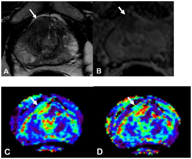
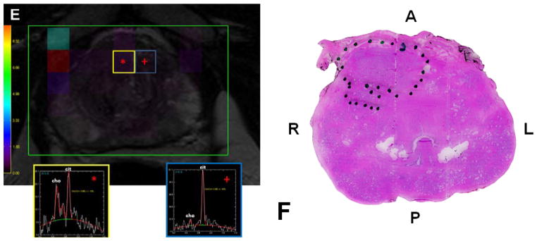
Purpose: We determined the prostate cancer detection rate of multiparametric magnetic resonance imaging at 3T. Precise one-to-one histopathological correlation with magnetic resonance imaging was possible using prostate magnetic resonance imaging based custom printed specimen molds after radical prostatectomy.
Materials and methods: This institutional review board approved prospective study included 45 patients (mean age 60.2 years, range 49 to 75) with a mean prostate specific antigen of 6.37 ng/ml (range 2.3 to 23.7) who had biopsy proven prostate cancer (mean Gleason score of 6.7, range 6 to 9). Before prostatectomy all patients underwent prostate magnetic resonance imaging using endorectal and surface coils on a 3T scanner, which included triplane T2-weighted magnetic resonance imaging, apparent diffusion coefficient maps of diffusion weighted magnetic resonance imaging, dynamic contrast enhanced magnetic resonance imaging and spectroscopy. The prostate specimen was whole mount sectioned in a customized mold, allowing geometric alignment to magnetic resonance imaging. Tumors were mapped on magnetic resonance imaging and histopathology. Sensitivity, specificity, positive predictive value and negative predictive value of magnetic resonance imaging for cancer detection were calculated. In addition, the effects of tumor size and Gleason score on the sensitivity of multiparametric magnetic resonance imaging were evaluated.
Results: The positive predictive value of multiparametric magnetic resonance imaging to detect prostate cancer was 98%, 98% and 100% in the overall prostate, peripheral zone and central gland, respectively. The sensitivity of magnetic resonance imaging sequences was higher for tumors larger than 5 mm in diameter as well as for those with higher Gleason scores (greater than 7, p <0.05).
Conclusions: Prostate magnetic resonance imaging at 3T allows for the detection of prostate cancer. A multiparametric approach increases the predictive power of magnetic resonance imaging for diagnosis. In this study accurate correlation between multiparametric magnetic resonance imaging and histopathology was obtained by the patient specific, magnetic resonance imaging based mold technique.
Copyright © 2011 American Urological Association Education and Research, Inc. Published by Elsevier Inc. All rights reserved.
Figures


Comment in
-
Use of magnetic resonance imaging to accurately detect and stage prostate cancer: the hype and the hope.J Urol. 2011 Nov;186(5):1756-7. doi: 10.1016/j.juro.2011.08.021. Epub 2011 Sep 23. J Urol. 2011. PMID: 21944134 No abstract available.
References
-
- American Cancer Society. Cancer Facts & Figures 2010. Atlanta: American Cancer Society; 2010.
-
- Kim CK, Park BK, Kim B. Localization of prostate cancer using 3T MRI: comparison of T2-weighted and dynamic contrast-enhanced imaging. J Comput Assist Tomogr. 2006;30:7–11. - PubMed
-
- Torricelli P, Cinquantini F, Ligabue G, Bianchi G, Sighinolfi P, Romagnoli R. Comparative evaluation between external phased array coil at 3 T and endorectal coil at 1.5 T: preliminary results. J Comput Assist Tomogr. 2006;30:355–61. - PubMed
-
- Park BK, Kim B, Kim CK, Lee HM, Kwon GY. Comparison of phased-array 3.0-T and endorectal 1.5-T magnetic resonance imaging in the evaluation of local staging accuracy for prostate cancer. J Comput Assist Tomogr. 2007;31:534–8. - PubMed
-
- Fütterer JJ, Heijmink SW, Scheenen TW, et al. Prostate cancer localization with dynamic contrast-enhanced MR imaging and proton MR spectroscopic imaging. Radiology. 2006;241:449–58. - PubMed
Publication types
MeSH terms
Grants and funding
LinkOut - more resources
Full Text Sources
Other Literature Sources
Medical
