

The gain in brain: novel imaging techniques and multiplexed proteomic imaging of brain tissue ultrastructure
- PMID: 21944260
- PMCID: PMC3265692
- DOI: 10.1016/j.conb.2011.08.004
The gain in brain: novel imaging techniques and multiplexed proteomic imaging of brain tissue ultrastructure
Abstract
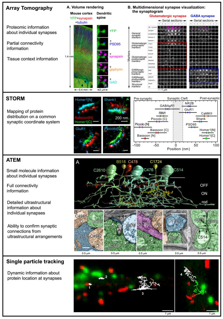
The rapid accumulation of neuroproteomics data in recent years has prompted the emergence of novel antibody-based imaging methods that aim to understand the anatomical and functional context of the multitude of identified proteins. The pioneering field of ultrastructural multiplexed proteomic imaging now includes a number of high resolution methods, such as array tomography, stimulated emission depletion microscopy, stochastic optical reconstruction microscopy and automated transmission electron microscopy, which allow a detailed molecular characterization of individual synapses and subsynaptic structures within brain tissues for the first time. While all of these methods still face considerable limitations, a combined complementary approach building on the respective strengths of each method is possible and will enable fascinating research into the proteomic diversity of the nervous system.
Copyright © 2011 Elsevier Ltd. All rights reserved.
Conflict of interest statement
KDM is cofounder of Aratome, LLC, a company working on the commercial application of array tomography, and holds a patent about array tomography technology.
Figures
Similar articles
-
Neuroproteomics - the tasks lying ahead.Electrophoresis. 2006 Jul;27(13):2819-29. doi: 10.1002/elps.200500892. Electrophoresis. 2006. PMID: 16739225 Review.
-
New observations in neuroscience using superresolution microscopy.J Neurosci. 2018 Oct 31;38(44):9459-9467. doi: 10.1523/JNEUROSCI.1678-18.2018. J Neurosci. 2018. PMID: 30381437 Free PMC article. Review.
-
Optical super-resolution microscopy in neurobiology.Curr Opin Neurobiol. 2012 Feb;22(1):86-93. doi: 10.1016/j.conb.2011.10.014. Epub 2011 Nov 1. Curr Opin Neurobiol. 2012. PMID: 22051692 Review.
-
Neuroproteomics Studies: Challenges and Updates.Methods Mol Biol. 2017;1598:3-19. doi: 10.1007/978-1-4939-6952-4_1. Methods Mol Biol. 2017. PMID: 28508355 Review.
-
Challenges of microtome-based serial block-face scanning electron microscopy in neuroscience.J Microsc. 2015 Aug;259(2):137-142. doi: 10.1111/jmi.12244. Epub 2015 Apr 23. J Microsc. 2015. PMID: 25907464 Free PMC article. Review.
Cited by
-
Building retinal connectomes.Curr Opin Neurobiol. 2012 Aug;22(4):568-74. doi: 10.1016/j.conb.2012.03.011. Epub 2012 Apr 11. Curr Opin Neurobiol. 2012. PMID: 22498714 Free PMC article. Review.
-
Retinal connectomics: towards complete, accurate networks.Prog Retin Eye Res. 2013 Nov;37:141-62. doi: 10.1016/j.preteyeres.2013.08.002. Epub 2013 Sep 7. Prog Retin Eye Res. 2013. PMID: 24016532 Free PMC article. Review.
-
Super-resolution microscopy for analyzing neuromuscular junctions and synapses.Neurosci Lett. 2020 Jan 10;715:134644. doi: 10.1016/j.neulet.2019.134644. Epub 2019 Nov 22. Neurosci Lett. 2020. PMID: 31765730 Free PMC article. Review.
-
Connecting connectomes.Neuroinformatics. 2013 Oct;11(4):389-92. doi: 10.1007/s12021-013-9207-0. Neuroinformatics. 2013. PMID: 24096724 Free PMC article. No abstract available.
-
A High-Resolution Method for Quantitative Molecular Analysis of Functionally Characterized Individual Synapses.Cell Rep. 2020 Jul 28;32(4):107968. doi: 10.1016/j.celrep.2020.107968. Cell Rep. 2020. PMID: 32726631 Free PMC article.
References
-
- Masland RH. Neuronal diversity in the retina. Curr Opin Neurobiol. 2001;11 :431–436. - PubMed
-
- Takamori S, Holt M, Stenius K, Lemke EA, Grønborg M, Riedel D, Urlaub H, Schenck S, Brügger B, Ringler P, Müller SA, Rammner B, Gräter F, Hub JS, De Groot BL, Mieskes G, Moriyama Y, Klingauf J, Grubmüller H, Heuser J, Wieland F, Jahn R. Molecular anatomy of a trafficking organelle. Cell. 2006;127:831–46. - PubMed
-
- Cheng D, Hoogenraad CC, Rush J, Ramm E, Schlager MA, Duong DM, Xu P, Wijayawardana SR, Hanfelt J, Nakagawa T, Sheng M, Peng J. Relative and absolute quantification of postsynaptic density proteome isolated from rat forebrain and cerebellum. Mol Cell Proteomics. 2006;5:1158–70. - PubMed
-
- Collins MO, Husi H, Yu L, Brandon JM, Anderson CN, Blackstock WP, Choudhary JS, Grant SG. Molecular characterization and comparison of the components and multiprotein complexes in the postsynaptic proteome. J Neurochem. 2006;97 (Suppl 1):16–23. - PubMed
Publication types
MeSH terms
Grants and funding
LinkOut - more resources
Full Text Sources
Medical
