

Reflectance confocal microscopy for the diagnosis of eosinophilic esophagitis: a pilot study conducted on biopsy specimens
- PMID: 21944314
- PMCID: PMC3425354
- DOI: 10.1016/j.gie.2011.07.020
Reflectance confocal microscopy for the diagnosis of eosinophilic esophagitis: a pilot study conducted on biopsy specimens
Abstract
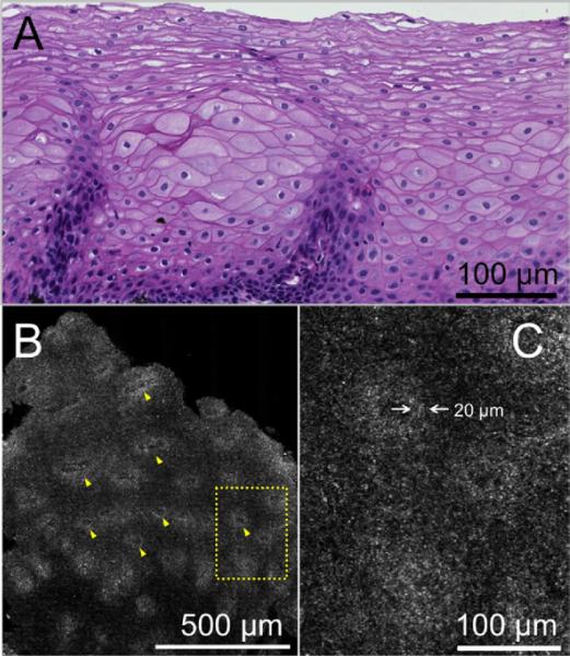
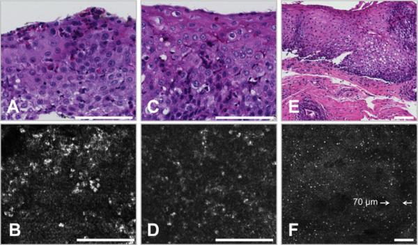
Background: Diagnosis of eosinophilic esophagitis (EoE) currently requires endoscopic biopsy and histopathologic analysis of the biopsy specimens to count intraepithelial eosinophils. Reflectance confocal microscopy (RCM) is an endomicroscopy technology that is capable of obtaining high-resolution, optically sectioned images of esophageal mucosa without the administration of exogenous contrast.
Objective: In this study, we investigated the capability of a high-speed form of RCM, termed spectrally encoded confocal microscopy (SECM), to count intraepithelial esophageal eosinophils and characterize other microscopic findings of EoE.
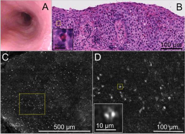
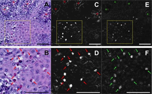
Design: A total of 43 biopsy samples from 35 pediatric patients and 8 biopsy samples from 8 adult patients undergoing EGD for EoE were imaged by SECM immediately after their removal and then processed for routine histopathology. Two SECM readers, trained on adult cases, prospectively counted intraepithelial eosinophils and detected the presence of abscess, degranulation, and basal cell hyperplasia on SECM images from the pediatric patients. A pathologist blinded to the SECM data analyzed the same from corresponding slides.
Setting: The Gastrointestinal Unit, Massachusetts General Hospital.
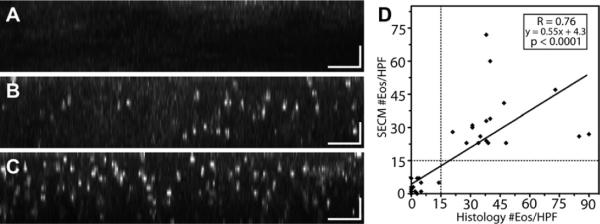
Results: Eosinophils by SECM demonstrated a higher reflectance than the surrounding cells and other inflammatory cells. There was good correlation between SECM and histology maximum eosinophil counts/high-power field (R = 0.76, P < .0001). Intra- and interobserver correlations for SECM counts were very good (R = 0.93 and R = 0.92, respectively; P < .0001). For the commonly used eosinophil count cutoff of 15 per high-power field, the sensitivity and specificity of SECM for EoE were 100%. The sensitivity and specificity for abscess, degranulation, and basal cell hyperplasia were 100% and 82%, 91% and 60%, and 94% and 80%, respectively. Intra- and interobserver agreements for these microscopic features of EoE were very good (κ = 0.9/0.9, 0.84/1.0, 0.91/0.81, respectively).
Limitation: Ex vivo study.
Conclusions: This study demonstrates that RCM can be used to accurately count intraepithelial eosinophils and identify other microscopic abnormalities associated with EoE on freshly excised biopsy samples. These findings suggest that RCM may be developed into a tool for assessing eosinophilic infiltration in the esophagus in vivo.
Copyright © 2011 American Society for Gastrointestinal Endoscopy. Published by Mosby, Inc. All rights reserved.
Figures
References
-
- Chehade M, Sampson HA. Epidemiology and etiology of eosinophilc esophagitis. Gastrointest Endosc Clin N Am. 2008;18:33–44. viii. - PubMed
-
- Kapel RC, Miller JK, Torres C, et al. Eosinophilic esophagitis: a prevalent disease in the United States that affects all age groups. Gastroenterology. 2008;134:1316–21. - PubMed
-
- Veerappan GR, Perry JL, Duncan TJ, et al. Prevalence of eosinophilic esophagitis in an adult population undergoing upper endoscopy: a prospective study. Clin Gastroenterol Hepatol. 2009;7:420–6. 426, e1–2. - PubMed
-
- Liacouras CA. Eosinophilic esophagitis. Gastroenterol Clin North Am. 2008;37:989–98. xi. - PubMed
-
- Atkins D, Kramer R, Capocelli K, et al. Eosinophilic esophagitis: the newest esophageal inflammatory disease. Nat Rev Gastroenterol Hepatol. 2009;6:267–78. - PubMed
Publication types
MeSH terms
Grants and funding
LinkOut - more resources
Full Text Sources
Medical
