Review
. 2011 Oct;42(4):585-601, ix.
doi: 10.1016/j.ocl.2011.07.004.

Emerging technologies for molecular therapy for intervertebral disk degeneration

Affiliations
Review

Emerging technologies for molecular therapy for intervertebral disk degeneration

Won C Bae et al. Orthop Clin North Am. 2011 Oct.

Abstract

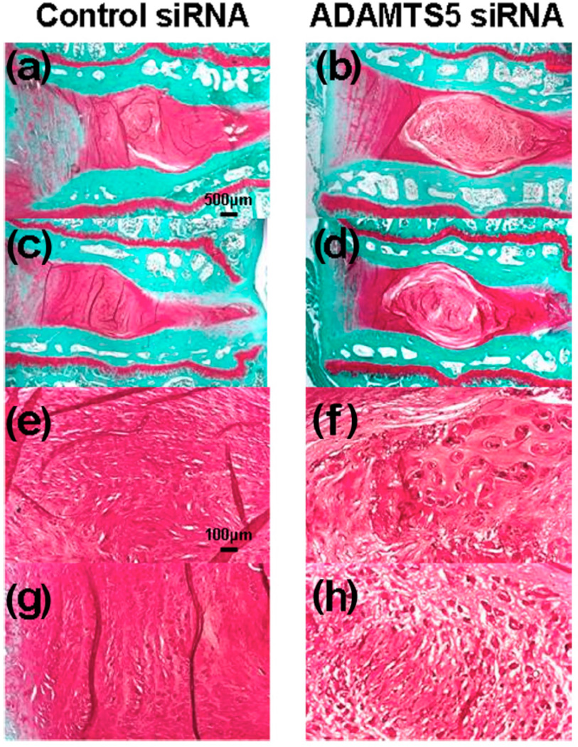
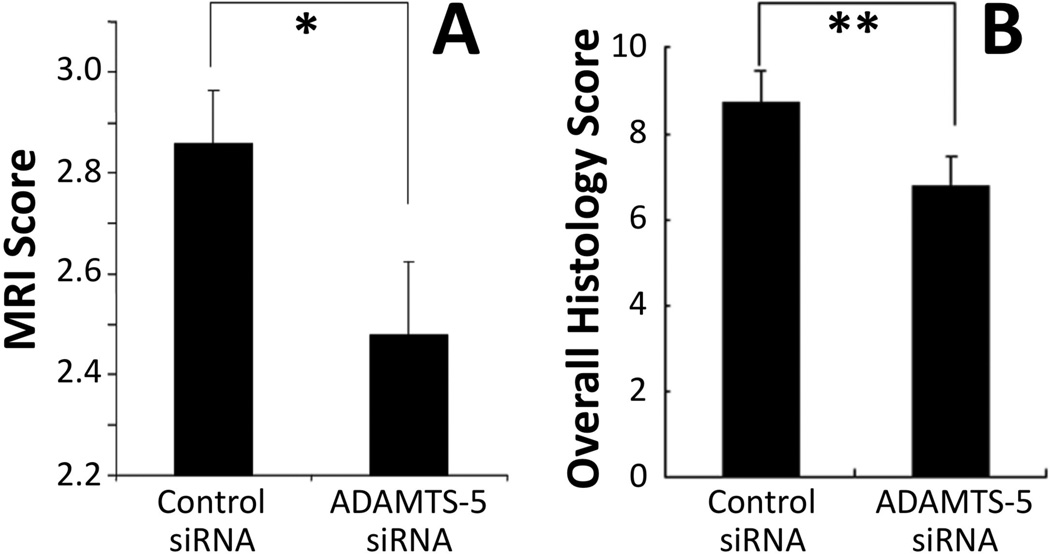
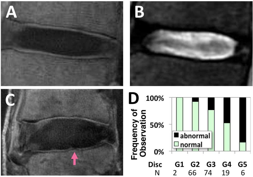
Intervertebral disks are biologically regulated by the maintenance of a balance between the anabolic and catabolic activities of disk cells. Therapeutic agents, initially evaluated using in vitro studies on disk cells and explants, have been used as intradiscal injections in preclinical settings to test in vivo efficacy. These include anabolic growth factors, other biostimulatory agents, and antagonistic agents against matrix-degrading enzymes and cytokines. Additional work is needed to identify patient populations, using methods such as MRI, and to better understand the mechanism of healing. Clinical trials are underway for a few of these agents and other promising candidates are on the horizon.


Figures

Figure 1
Figure 1. Changes in the intervertebral disc (IVD) height index (DHI) after anular puncture and recombinant human growth and differentiation factor-5 (rhGDF-5) injection
In a needle-puncture model of disc degeneration (4 weeks) in adolescent (5–6 months-old) rabbits, a single injection of rhGDF-5 restored disc height (A). In a study using 2-year old rabbits, rhGDF-5 disc height was also effectively recovered (B). (Modified from Chujo T, et al. Effects of growth differentiation factor-5 on the intervertebral disc- in vitro bovine study and in vivo rabbit disc degeneration model study. Spine 2006;31(25):2909-17 and Chujo T, et al. In vivo effects of recombinant human growth and differentiation factor-5 on the repair of the mature rabbit intervertebral disc. Spine J, 6(5):23S–24S, 2006 (North American Spine Society, 21st Annual Meeting Proceeding, abstract). AF: annulus fibrosus; PBS: phosphate-buffered saline.
Figure 2
Figure 2. Quantitative magnetic resonance (MR) parameter maps of thrombin-induced degraded rabbit lumbar spines after injection with the growth factor, recombinant human growth and differentiation factor-5 (rhGDF-5)
Four weeks after thrombin injection (100 U/10 µl) into adolescent rabbit discs, a single injection of saline (10 µl) (A, B) or rhGDF-5 (10 µg/10 µl) (C, D) was given. T2 (A, C) and T1rho (B, D) MRI maps were obtained 12 weeks later. High MR values in the nucleus pulposus regions are apparent in the samples treated with the growth factor (C, D). (Modified from Bae WC, et al. Effect of rhGDF-5 on the thrombin model of rabbit intervertebral disc degeneration: T1rho quantification using 3T MRI. Rad Soc North Am 2009;95:SSE14-02, abstract)
Figure 3
Figure 3. Treatment with recombinant human growth and differentiation factor-5 (rhGDF-5) restored T2 and T1rho values after thrombin-induced degeneration in adolescent rabbit discs
Four weeks after thrombin injection (100 U/10 µl) into adolescent rabbit discs, a single injection of saline (10 µl) or rhGDF-5 (10 µg/10 µl) was given. T2 and T1rho MRI maps were obtained 12 weeks later. Regions of interest were selected to determine T2 and T1rho values in the nucleus pulposus (NP). Both T2 (A) and T1rho (B) values of the NP were higher in the rhGDF-5-treated group, approaching those of the unoperated group. The saline group had significantly lower values compared to the unoperated control. (Modified from Bae WC, et al. Effect of rhGDF-5 on the thrombin model of rabbit intervertebral disc degeneration: T1rho quantification using 3T MRI. Rad Soc North Am 2009;95:SSE14-02, abstract)
Figure 4
Figure 4. Treatment with recombinant human growth and differentiation factor-5 (rhGDF-5) inhibits the expression of molecules related to disc degradation, pain, and neovascularization in thrombin-degenerated adolescent rabbit discs
Four weeks after thrombin injection (100 U/10 µl) into adolescent rabbit discs, a single injection of saline (10 µl) or rhGDF-5 (10 µg/10 µl) was given. Twelve weeks later, mRNA expression levels for ADAMTS-5 (A), ADAMTS-4 (B), COX-2 (C) and VEGF (D), in saline-and rhGDF-5-treated discs showed a trend or significantly lower levels in the rhGDF-5-treated samples. (Modified from Masuda K, et al. Intradiscal injection of recombinant human growth and differentiation factor-5 significantly suppressed the expression of cytokines, catabolic enzymes and pain markers in the rabbit anular puncture model. Paper presented at: The 35th Annual Meeting of the International Society for the Study of the Lumbar Spine 2009:47, abstract) ADAMTS: a disintegrin and metalloproteinase with a thrombospondin type 1 motif; COX-2: cyclooxygenase-2; VEGF: vascular endothelial growth factor.
Figure 5
Figure 5. Histology of anular puncture-induced degenerated adolescent rabbit discs injected with control small interference RNA (siRNA) or ADAMTS-5 siRNA
Representative safranin-O-stained sections after injection of ADAMTS-5 siRNA or control siRNA in the rabbit anular puncture model of disc degeneration. Eight weeks after the siRNA injections, the control siRNA group displayed a complete loss of nucleus pulposus (NP) tissues that had been replaced by a fibrocartilaginous tissue (a, c). The severely degenerated discs that had received the control siRNA showed a loss of proteoglycans and the collapsed, wavy fibrocartilage lamellae typical of the anulus fibrosus (AF), with associated fibrochondrocytes (e, g). In the ADAMTS5 siRNA-injected discs, safranin-O staining demonstrated the maintenance of intervertebral disc structure with a lightly stained matrix and large cells (b, d); the NP was rounded and bloated in appearance, and consisted of numerous large, vacuolated cells and smaller chondrocyte-like cells (f, h). A clear demarcation was seen between the NP and inner anulus in the ADAMTS5 siRNA-injected discs. (Magnification is 20× (a–d) or 100× (e–h)). The level in a, b, e, and f is L2/3, and in c, d, g, and h is L4/5. (Reproduced from Seki S, et al. Effect of small interference RNA (siRNA) for ADAMTS5 on intervertebral disc degeneration in the rabbit anular needle-puncture model. Arthritis Research & Therapy 2009;11:R166). ADAMTS: a disintegrin and metalloproteinase with a thrombospondin type 1 motif.
Figure 6
Figure 6. Magnetic resonance imaging (MRI) and histology grading scores of anular puncture-induced degenerated adolescent rabbit discs injected with control small interference RNA (siRNA) or ADAMTS-5 siRNA
(A) MRI assessment 8 weeks after siRNA injection, using a modified Thompson scale (1–4). The MRI grade in the ADAMTS-5 siRNA-treated discs show significantly better (lower) MRI grade compared to control siRNA-treated discs. (B) Histological assessment for structure, cellularity and matrix staining showed significantly better (lower) overall score (range 0–12) for ADAMTS-5 siRNA-treated samples. Mean ± S.E.M., n=6. * p<0.05, ** p<0.01 Mann-Whitney U test. (Modified from Seki S, et al. Effect of small interference RNA (siRNA) for ADAMTS5 on intervertebral disc degeneration in the rabbit anular needle-puncture model. Arthritis Research & Therapy 2009;11:R166).
Figure 7
Figure 7. Retention of radiolabeled bone morphogenetic-7 (BMP-7) in a rabbit disc
Normal rabbits that received a single intradiscal injection of 125I-labeled BMP-7 (otherwise known as osteogenic protein-1) were imaged using autoradiography (A) 6 hours, (B) 24 hours or (C) 28 days after the injection. The signal from the radiolabeled BMP-7 is prominent even after 14 days. (Modified from Pierce, A. et al. Distribution, pharmacokinetics and excretion of 125-Iodine labeled BMP-7 (OP-1) following a single dose administration in lumbar IVD or knee joint of NZW rabbits. Paper presented at: The sixth International conference on bone morphogenetic protein 2006; Cavtat, Croatia.)
Figure 8
Figure 8. Evaluation of region of cartilage endplate using ultrashort time-to-echo (UTE) magnetic resonance imaging (MRI)
A normal lumbar disc imaged using UTE MRI (A) and conventional T2-weighted spin echo MRI (B). Note the characteristic high-intensity lines (A) near the regions of cartilage endplate. The same regions appear dark in conventional MRI (B). Abnormalities such as focal signal loss have been observed (arrow, C) and correlated with the grade of the adjacent disc (D). (Modified from Bae WC, et al. Ultrashort time-to-echo MRI of human intervertebral disc endplate: association with disc degeneration. Proc Int'l Soc Magn Reson Med 2010;18:534)

References

    1. Maeda S, Kokubun S. Changes with age in proteoglycan synthesis in cells cultured in vitro from the inner and outer rabbit annulus fibrosus. Responses to interleukin-1 and interleukin-1 receptor antagonist protein. Spine. 2000;25:166. - PubMed
    1. Antoniou J, Steffen T, Nelson F, et al. The human lumbar intervertebral disc: evidence for changes in the biosynthesis and denaturation of the extracellular matrix with growth, maturation, ageing, and degeneration. J Clin Invest. 1996;98:996. - PMC - PubMed
    1. Buckwalter JA, Kuettner KE, Thonar EJ. Age-related changes in articular cartilage proteoglycans: electron microscopic studies. J Orthop Res. 1985;3:251. - PubMed
    1. Buckwalter JA, Pedrini MA, Pedrini V, et al. Proteoglycans of human infant intervertebral disc. Electron microscopic and biochemical studies. J Bone Joint Surg [Am] 1985;67:284. - PubMed
    1. Cole TC, Ghosh P, Taylor TK. Variations of the proteoglycans of the canine intervertebral disc with ageing. Biochim Biophys Acta. 1986;880:209. - PubMed

Publication types

MeSH terms